Editorial - Volume 2 - Issue 5
The Uncancerous Helix-Benign Fibrous Histiocytoma
Anubha Bajaj*
*Consultant Histopathology, A.B. Diagnostics, New Delhi, India
Received Date : July 19, 2022
Accepted Date : Aug 19, 2022
Published Date: Sep 05, 2022
Copyright:© Anubha Bajaj 2022
*Corresponding Author : Anubha Bajaj, Consultant Histopathology, A.B.Diagnostics, New Delhi, India.
Email:anubha.bajaj1@outlook.com
DOI: Doi.org/10.55920/2771-019X/1234
Introduction
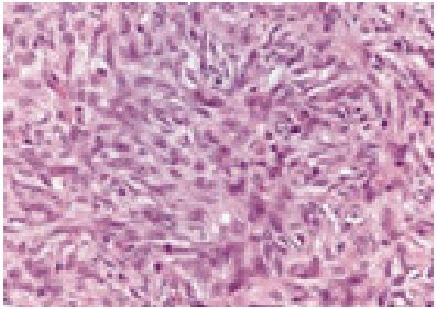
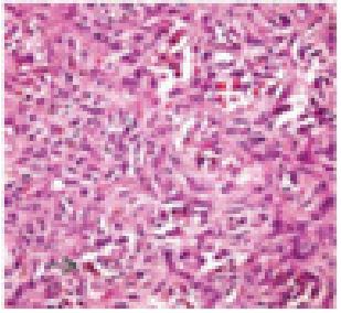
Benign fibrous histiocytoma occurs as an exceptional, painless, subcutaneous, mesenchymal, fibrohistiocytic neoplasm devoid of dermal incrimination. Additionally designated as deep dermatofibroma, deep seated benign fibrous histiocytoma commonly emerges within subcutaneous tissue, deep seated soft tissue or parenchymal organs. Generally, benign fibrous histiocytoma arises in adult males > 25 years and appears between 6 years to 84 years with a median age of disease emergence at 40 years. A male predominance is observed. Commonly, benign fibrous histiocytoma emerges within lower or upper extremities, head and neck or trunk. Neoplasms confined to deep soft tissue of retroperitoneum, mediastinum or pelvis are infrequently discerned [1,2]. Deep seated benign fibrous histiocytoma emerges as a painless, gradually progressive, well circumscribed or encapsulated tumefaction [1,2]. Grossly, firm, yellow to tan, well circumscribed tumefaction encompassed with a pseudo-capsule is discerned. Pseudo-capsule may be absent where tumefaction extends to the cutis [1,2]. Characteristically, tumour magnitude is ~ 4 centimetres and varies from 0.5 centimetres to 25 centimetres. Cut surface exhibits variable foci of haemorrhage [1,2]. Upon microscopy, neoplasm exhibits a prominent storiform pattern configured of uniform, spindle-shaped cells imbued with eosinophilic cytoplasm with ill defined cellular perimeter and bland, elongated or plump nuclei with vesicular chromatin and delicate nucleoli. Tumour nuclei are devoid of nuclear atypia [1,2]. tumefaction is admixed with disseminated lymphocytes, plasma cells multinucleated giant cells, osteoclastic giant cells or foamy macrophages [1,2]. Frequently, prominent vascular articulations may configure a hemangiopericytoma-like pattern. Mitotic figures appear ≤5 mitosis per 10 high power fields [1,2]. Intervening stroma is myxoid or hyaline. Focal necrosis or tumour infiltration of lymphatic and vascular configurations is exceptional. Tumour perimeter is non-infiltrative and peripheral entrapment of adipose tissue is absent. Focal entrapment of collagen fibres may occur [1,2].
Figure 1: Benign fibrous histiocytoma depicting whorls and storiform pattern of spindle shaped cells demonstrating uniform, bland nuclei and vascular prominence [5].
Figure 2: Benign fibrous histiocytoma delineating whorls and fascicles of spindle shaped cells with a focal storiform pattern, uniform, vesicular nuclei and red cell extravasation [6].
Benign fibrous histiocytoma is immune reactive to CD34 or smooth muscle actin [3,4]. Benign fibrous histiocytoma is immune non reactive to keratin, desmin, EMA, S100 protein, α1-antitrypsin, α1-antichymotrypsin, CD117, CD31, CD68, carcinoembryonic antigen (CEA), factor VIII, Ki-67 protein, Leu7, and vimentin [3,4]. Benign fibrous histiocytoma requires segregation from neoplasms such as dermatofibrosarcoma protuberans, nodular fasciitis, solitary fibrous tumour, fibroma, fibro-sarcoma, neurofibroma or Kaposi’s sarcoma. Comprehensive surgical extermination of the neoplasm is an optimal mode of therapy. Inadequate surgical eradication is associated with tumour reoccurrence [3,4]. Localized tumour reoccurrence is commonly discerned. Distant metastasis in undocumented and tumour associated mortality is extremely exceptional [3,4].
References
- Kirschnick LB, Schuch LF, et al. Benign fibrous histiocytoma of the oral and maxillofacial region: A systematic review. Oral Surg Oral Med Oral Pathol Oral Radiol. 2022; 133(2): e43-e56.
- Wang H, Zhang S, et al. Successful treatment of recurrent benign fibrous histiocytoma by complete surgical removal and appropriate ocular surface reconstruction: A case report. Medicine (Baltimore). 2020; 99(44): e22941.
- Popescu N, Gheorghe GA et al. Conjunctival benign fibrous histiocytoma in a 57-year-old man. Rom J Ophthalmol. 2019; 63: 273-6.
- Nguyen A, Vaudreuil A et al. Clinical Features and Treatment of Fibrous Histiocytomas of the Tongue: A Systematic Review. Int Arch Otorhinolaryngol. 2018; 22(1): 94-102.
- Image 1 Courtesy: Pathology outlines
- Image 2 Courtesy: Basic medical key


