Clinical Image - Volume 2 - Issue 5
Intrathoracic hepatic liver as a cause of chronic dyspnea and thoracic pain
Víctor Hugo Alcalá Torres1; Marian Eliza Izaguirre-Pérez2*; Luis Osvaldo Suárez Carreón3; Dennis Salas Gradilla4; Gilberto González Pérez1; Juan Ignacio Mandujano-Sánchez5
1Laparoscopic surgery / Civil Hospital Dr. Juan I. Menchaca / Guadalajara, Jalisco, México.
2General Surgery / Regional Hospital Dr.Valentín Gómez Farías, Institute of Security and Social Services for State Workers (ISSSTE) / Zapopan, Jalisco, México.
3General Surgery / Western National Medical Center, Mexican Social Security Institute (IMSS) / Guadalajara, Jalisco, México.
4Plastic and reconstructive surgery / Civil Hospital Fray Antonio Alcalde / Guadalajara, Jalisco, México.
5Trauma surgery / Lomas Verdes Hospital, Mexican Social Security Institute (IMSS) / Naucalpan de Juárez, Edo. México, México.
Received Date : Aug 15, 2022
Accepted Date : Sep 19, 2022
Published Date: Sep 27, 2022
Copyright:© Marian Eliza IzaguirrePérez 2022
*Corresponding Author : Marian Eliza Izaguirre-Pérez, General Surgeon Av. Washington 4957, Zapopan, Jalisco. CP 45027.Tel: 3336736707
Email:marian_eliza@hotmail.es
DOI: Doi.org/10.55920/2771-019X/1252
Abstract
The presentation of ectopic supradiaphragmatic liver tissue is remarkably rare. We present the case of a 40-y-o female patient. With 6-mo of dyspnea with chest and abdominal pain. Computed tomography revealed a well-circumscribed homogenous mass. A thoracoscopic approach revealed an intrathoracic ectopic hepatic parenchyma.
Description
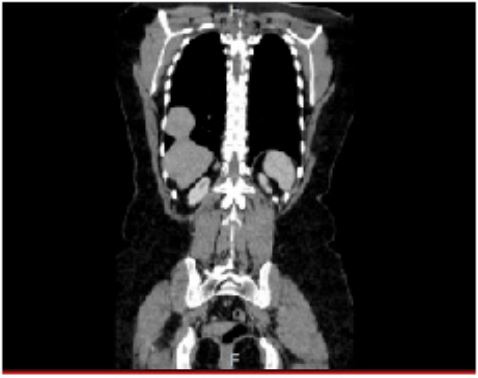
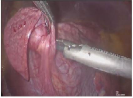
A 40-year-old Hispanic female was referred to our center complaining of 6-mo episodes of dyspnea associated with chest pain and abdominal pain located in the right hypochondrium refractory to medical treatment. She denied any previous history of trauma or surgery. There were no positive findings on the physical examination. Computed tomography (CT) revealed a well-circumscribed homogenous soft tissue mass, described as an apparent protrusion of the liver into the thorax of 6 cm, suggesting a diaphragmatic hernia (Figure 1). A right chest wall thoracoscopic was performed, and the operative findings revealed intrathoracic ectopic hepatic parenchyma of 3.5 cm x 2 cm x 1.5 cm, with no evidence of a hernial defect (Figure 2).
Ectopic tissue was resected with an Endo-GIA staple instrument and subsequently diaphragmatic plication and reinforcement of the stapling line. A Pneumokit was placed to prevent the formation of a pneumothorax. The patient had a good evolution after the surgery, a chest X-ray was taken 24 hours after surgery, showing adequate pulmonary expansion. She was discharged 2 days after surgery with no complications. The pathologic report showed ectopic liver with fibrosis, chronic inflammation, and hemorrhage.
Ectopic supradiaphragmatic liver tissue is a rare entity and may sometimes go unnoticed on a radiological examination. This pathology should be considered in symptomatic patients with chronic pain and dyspnea or when diagnosing asymptomatic masses with a supradiaphragmatic location.
Figure 1: Computed tomography shows a lesion of about 4 cm in the coronal plane.
Figure 2: Video-assisted thoracoscopy with the presence of ectopic intrathoracic liver tissue and partial diaphragm paralysis.


