Clinical Image - Volume 2 - Issue 5
Intraosseous bubble or gas: Emphysematous osteomyelitis
Amarkak Abdillahi Wais; Soufiane Rostoum*; Daoud Ali Mohamed; Onka Behyamet; JEl Fenni; Hassan EnNouali; Rachida Saouab
*Radiology Department, Mohamed V military hospital, Rabat, Morocco.
Received Date : Aug 23, 2022
Accepted Date : Sep 23, 2022
Published Date: Oct 03, 2022
Copyright:© Soufiane Rostoum 2022
*Corresponding Author : Soufiane Rostoum, Radiology Department,Mohamed V military hospital, Rabat, Morocco.Tel: +212649496021
Email:rostoum.soufiane@gmail.com
DOI: Doi.org/10.55920/2771-019X/1256
Clinical presentation
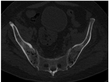
60-year-old diabetic patient, followed for Hodgkin's lymphoma who presented to the emergency room for diffuse abdominal pain in a febrile context. The biological examination showed hyperleucocytosis with a high CRP (320mg/l). An ultrasound was performed showing a voluminous lateralized right pelvic collection confirmed by an abdominal CT scan. The patient benefited from a percutaneous drainage objectifying a purulent liquid. The cytobacteriological examination came back in favor of E.coli. The patient was put on ceftriaxone-based antibiotic therapy. Evolution is marked by a decrease in fever with persistence of high CRP. A CT scan showed a clear regression of the collection with the appearance of intraosseous air bubbles in the iliac wings without cortical involvement, suggesting emphysematous osteomyelitis (Figure 1).
Figure 1: Axial pelvic computed tomography (bone window) showing multiple small bubbles or gas in the intramedullary iliac bone without cortical destruction.
Discussion
The intraosseous bubble or gas is a rare phenomenon, a sign first described in 1981 by P C Ram et al. as a sign of osteomyelitis [1]. When intraosseous gas is seen in the extra-axial skeleton, this sign is pathognomonic for emphysematous osteomyelitis (OE) [1,2]. The most commonly affected sites are the vertebrae, pelvis, sacrum, femur, tibia, fibula, and midfoot [2,3]. Emphysematous osteomyelitis is usually a clinically unsuspected condition that is diagnosed based on radiological findings. Most often, it affects patients with an underlying comorbidity: Diabetes and malignant tumors are the most common risk factors. Other incriminated factors are Crohn's disease, alcohol abuse, prolonged corticosteroid therapy, etc [2,4]. Computed tomography (CT) is the imaging modality of choice because it is the most sensitive method for detecting intraosseous gas [1,2]. Typically, emphysematous osteomyelitis is seen as multiple small and irregular foci of intramedullary gas, without cortical destruction, a key diagnostic feature [1]. The combination of intraosseous and extraosseous gases without cortical destruction is very specific to this entity [2,4]. The germs most incriminated at the origin of EO are the members of the family of enterobactericiacea or a particular anaerobic Fusobacterium necrophorum. Various other organisms that have been described as causing emphysematous osteomyelitis are Klebsiella pneumoniae, Enterobacter aerogenes, Clostridium spp, Staphylococcus aureus, non-hemolytic streptococci and enterococci, and Pseudomonas spp [2,3].
In the absence of a history of surgery, trauma, or degenerative change, bone lymphangiomatosis, osteonecrosis and neoplasm, the intraosseous gas is highly suggestive of emphysematous osteomyelitis. However, intraosseous gas in vertebral bodies is generally due to a non-infectious cause, most commonly a degenerative process [2,5].
Treatment of patients with emphysematous osteomyelitis should be aggressive with long-term antibiotic therapy (at least 4 weeks). Surgery should be reserved for complications such as abscess formation or necrosis, and for patients who do not respond to antimicrobial agents [2,5].
Conflict of Interests
All authors have no conflict of interest to declare.
Funding
All authors have no funding source to declare.
Ethical Approval
Consent Written Consent Has Been Obtained.
References
- Ram P, Martinez S, Korobkin M, Breiman R, Gallis H & Harrelson J. CT detection of intraosseous gas: a new sign of osteomyelitis. American Journal of Roentgenology, 1981; 137(4): 721–723.
- Mujer MT, Rai MP, Hassanein M, Mitra S. Emphysematous Osteomyelitis. BMJ Case Rep. 2018 Jul 15; 2018: bcr2018225144.
- Lee J, Jeong CH, Lee MH, et al. Emphysematous Osteomyelitis due to Escherichia coli. Infect Chemother 2017; 49: 151–4.
- Larsen J, Mühlbauer J, Wigger T, Bardosi A. Emphysematous osteomyelitis. Lancet Infect Dis. 2015 Apr; 15(4): 486. [PMID: 25809901 DOI: 10.1016/S1473-3099(14)71017-5].
- Huéscar LL, San Miguel J, Espinosa LMMB, García P, Benedito CRS, Ordás PO & Emergency CT. Intraosseous air bubbles, a sign of emphysematous osteomyelitis.


