Clinical Image - Volume 2 - Issue 5
Ultrasound “whirlpool sign” of small bowel volvulus
Benmoussa Meryem*; El Haddad Siham; El Allali Nazik; Chat Latifa
Departement of radiology, Children’s Hospital of Rabat, Morocco
Received Date : Aug 28, 2022
Accepted Date : Sep 27, 2022
Published Date: Oct 07, 2022
Copyright:© Benmoussa Meryem 2022
*Corresponding Author : Benmoussa Meryem, Departement of radiology, Children’s Hospital of Rabat, Morocco.
Email:benmoussa.mer93@gmail.com
DOI: Doi.org/10.55920/2771-019X/1259
Keywords
Volvulus; Whirlpool sign; Intestinal obstruction
Comment
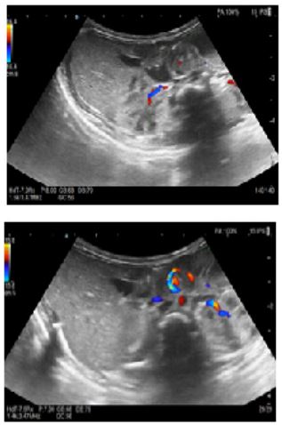
Small bowel volvulus is a real surgical emergency. Early diagnosis and prompt operation are essential to prevent gangrene in the small intestine, which is associated with high morbidity and mortality. This is a life-threatening complication of intestinal malrotations, which is defined by abnormal twisting of a loop of small bowel around the axis of its own mesentery during embryologic development [1]. Clinical signs are represented by the high neonatal obstruction syndrome. Imaging has an essential role in diagnosis. Abdominal X-Ray shows marked distension of the intestines concerning for acute obstruction, however it can further lead to missed or delayed diagnoses. The associated abdominal Doppler ultrasound confirm the diagnosis of volvulus by showing “whirlpool sign”. This sign corresponds to a clockwise wrapping of the superior mesenteric vein and the mesentery around the superior mesenteric artery.
Figure 1 and 2: Color Doppler ultrasound showing an epigastric pseudo-mass with “whirlpool sign”.
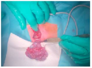
We report the case of a newborn at 11 days of life, admitted to the pediatric surgical emergency department of the Children's Hospital of Rabat for high neonatal obstruction syndrome with bilious vomiting and abdominal pain. Clinical examination found a flat abdomen. The abdominal X-ray found gastric distention with a dilated fluid-filled duodenum. The associated abdominal Doppler ultrasound shows the “whirlpool sign”, that confirms the diagnosis. (Figure 1 and 2). Surgical treatment consisted of detorsion of the volvulus with Ladd procedure and appendectomy. (Figure 3)
Figure 3: Perioperative image of small bowel volvulus on common mesentery
Authors' statements
The authors declared no potential conflicts of interest with respect to the research, authorship, and/or publication of this article.
References
- Acute small bowel volvulus in adults. A sporadic form of strangulating intestinal obstruction. Roggo A, Ottinger LW. https://www.ncbi.nlm.nih.gov/pmc/articles/PMC1242584/Ann Surg. 1992; 216: 135–141.


