Clinical Image - Volume 2 - Issue 5
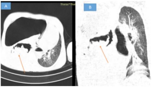
Water lily appearance synonymous with hydatid cyst
Diallo Ibrahima Dokal*; Traore Wend-Yam Mohamed; El Haddad Siham; Allali Nazik; Chat Latifa
Radiology Department of the Children’s Hospital of Rabat, CHU Ibn Sina of Rabat, Morocco.
Received Date : Aug 26, 2022
Accepted Date : Oct 01, 2022
Published Date: Oct 13, 2022
Copyright:© Diallo Ibrahima Doka 2022
*Corresponding Author : Diallo Ibrahima Dokal, Avenue Maghreb el araabi, Apt 16, immeuble 60, rabat, Marocco.Tel: +212681259433
Email: dialloibrahimadokal@gmail.com
DOI: Doi.org/10.55920/2771-019X/1263
The water lily appearance corresponds to the presence of wavy or serpiginous structures within an excavated mass. It indicates the presence of a complicated hydatid cyst. Hydatid cyst is a cosmopolitan anthropozoonosis, related to the development of a larval form of a parasite "echinococcus granulosis". The pulmonary localization comes in second place after the hepatic one [1]. From an anatomical point of view, a hydatid cyst appears to be made up of a reactionary tissue membrane belonging to the host and two other membranes, constituents of the parasite; one is acellular (anhistic cuticle) whereas the second, the germinative proliga, gives rise to daughter vesicles. It contains hydatid fluid in rock water containing numerous protoscolex and membrane debris known as hydatid sand [2]. It is suspected in the presence of hemoptysis, rejection of membranes, hydatid vomitus or non-specific respiratory symptoms concomitant with a particular context in this case when contact with dogs, which are the definitive hosts, is found. Its diagnosis is made possible by imaging through chest X-ray and thoracic CT, which are by far the most used means. Thus, different aspects of hydatid cysts are found on imaging, depending on the stage of evolution: it may be a simple cyst (unilocular cyst), identified as a low-toned, watery, well-limited, homogeneous opacity, known as a cannonball; corresponding to a mass of liquid density with a thin, regular wall on CT; or a complicated cyst (fissuring, rupture, compression). In the case of rupture, the hydatid cyst appears as a hydroaerobic image with an undulating border, describing serpentine structures, reminiscent of a "water lily" flower [1]. This sign is of great help in the etiological research in front of a hydro-pneumothorax of great abundance, thus implying a surgical management.
Declaration of Interests
The authors declare that they have no conflict of interests.

Figure 1: Axial (A) and coronal (B) parenchymal window CT sections showing compressive hydro-pneumothorax responsible for right lung collapse on rupture of a hydatid cyst. Note the wavy appearance of the proligeral membrane describing the water lily appearance (arrow).

