Case Report - Volume 2 - Issue 5
Solitary plasmacytoma of the sternum: A case report
Moudafia Z1*; Lamri.H1; Dahmani B1; Omari Tadlaoui. S1; Alaoui Rachidi S1,2
1Radiology Department, Tangier-Tetouan-Al-Hoceima University Hospital. Morocco.
2Faculty of Medicine and Pharmacy of Tangier, Abdelmalek Essaadi University, 90000 Tangier, Morocco.
Received Date : Aug 26, 2022
Accepted Date : Oct 04, 2022
Published Date: Oct 15, 2022
Copyright:© Moudafia Z 2022
*Corresponding Author : Moudafia Z, Radiology Department, TangierTetouan-Al-Hoceima University Hospital, (2) Faculty of Medicine and Pharmacy of Tangier, Abdelmalek Essaadi University, 90000 Tangier, Morocco
Email: zinmoud@gmail.com
DOI: Doi.org/10.55920/2771-019X/1265
Abstract
Costal solitary plasmacytoma is a rare entity, which should be evoked in front of a sternal tumor with intraosseous development on imaging. Radiotherapy is the most effective treatment modality for solitary plasmacytoma of the bone.The risk of progression to multiple myeloma conditions the prognosis, which justifies post-treatment monitoring and suggests a reflection on the exact place of early adjuvant chemotherapy. We report a case of a an old patient with a Sternal Plasmacytoma.
Keywords: Sternal Plasmacytoma; CT-scan; Protein immunoelectrophoresis; bone biopsy; radiotherapy.
Introduction
Solitary plasmacytoma of bone (osseous plasmacytoma) is a rare entity characterized by the proliferation of malignant plasma cells, These are localized to a bone segment without diffuse bone marrow invasion. The dorsolumbar location is the most frequent; sternal involvement is rarely described.
Case presentation
We report the case of a 58 year old patient with a bilateral inguinal hernia operated in 1987. He presented for 6 months a sternal bone swelling without local inflammatory signs. The biological check-up revealed a monoclonal peak at 6g/L. Protein immunoelectrophoresis (IEPP) associated with the determination of light chains: monoclonal IgG type IgG Kappa. Medullogram 3% Plasmocytes No anemia and No renal insufficiency and no hypercalcemia. X-rays of the lung, spine and skeletal bones were without abnormality.
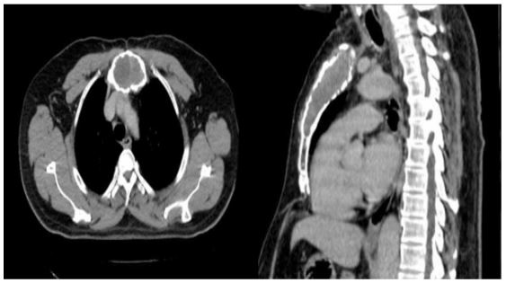
The patient underwent a CT scan which showed an osteolytic tumor process centered on the sternum with intraosseous development without soft tissue invasion measuring 38x54mm, with no other spinal, thoracic, or costal locations (Figure1). A scan-guided bone biopsy was performed, the anatomopathological examination of which concluded to a solitary plasmacytoma of the sternum. The treatment included radiotherapy alone, Indeed he received a dose of 50 Gy in 25 fractions of 2 Gy from 19/03/2019 to 30/04/2019 by 3D conformal radiotherapy technique. The evolution was a radiological stability, the EPP(Protein electrophoresis) peak at 3.9 g/L, the patient did not develop multiple myeloma or other plasma cell localizations

Figure 1: Axial CT sections and sagittal reconstructions showing osteolytic tumor process centered on the sternum with intraosseous development: STERNAL PLASMACYTOMA.
Discussion
Solitary plasmacytoma (SP) is a rare tumor, representing less than 5% of plasma cell neoplasia [1, 2]. It is defined by the presence of an isolated plasma cell tumor localized to a bone segment. It is a focal lesion composed of malignant plasma cells but without diffuse marrow invasion, which distinguishes it from multiple myeloma. It mainly affects the axial skeleton. Solitary plasmacytoma includes two distinct entities in relation to tumor location: either bone (intramedullary solitary plasmacytoma) or soft tissue (extramedullary solitary plasmacytoma) [3]. Solitary bone plasmacytoma is characterized by a single lesion most often affecting the axial skeleton, particularly the vertebrae. The bony or intramedullary form is the most common [4]. Men are most often affected, in about two thirds of cases, with an average age of 55 years, ten years younger than the age of onset of multiple myeloma [5]. Our patient is in the age range most frequently reported in the literature, namely between 50 and 60 years. The predominance of males is clear, with a sex ratio of 3 to 4. The involvement is costal, sternal, clavicular or scapular in about 20% of cases [5]. Our patient therefore presents a sternal location. It may be manifested by pain or bone swelling, as in our patient, or it may be an incidental radiological finding [7]. The diagnosis of solitary bone palsmacytoma implies the presence of a single bone lesion with biological and radiological investigations that did not reveal other locations [6]. In order to confirm the diagnosis, a complete general workup is necessary to rule out multiple myeloma. A number of criteria are necessary to make the diagnosis: absence of anemia, normal phosphocalcium status, normal renal function, normal bone marrow cytology in at least two different sites, and absence of other lesions on skeletal radiographs or CT or MRI scans [9,10]. The biological workup and radiographs of our patient were without abnormality. Standard radiography shows a well-limited bone gap without peripheral sclerosis, which may lead to bone destruction with soft tissue invasion [10,11,12]. CT scan shows a lytic lesion taking up contrast intensely and homogeneously. It allows a better study of the local extension and searches for possible infraradiological bone localizations [13,12].
Our patient presented on his CT scan a heterodense osteolytic formation centered on the sternum and with intraosseous development without invasion of the soft parts. Magnetic resonance imaging typically reveals a hypointense lesion in T1, T2 and after gadolinium injection [5,9]. The diagnosis of certainty is based on the anatomopathological study of the surgical specimen or scanned trans-parietal biopsies, showing a sheet-like plasma cell proliferation with dysmorphic plasma cells with nuclear abnormalities and normal cytoplasm. Immunohistochemistry confirmed the monoclonal nature of the tumor cells by demonstrating intracytoplasmic immunoglobulins. The scan-guided biopsy performed for our patient confirmed the diagnosis. The treatment of OSP is mainly based on radiotherapy (RT); these are radiosensitive and radiocurable tumors [ 14,15 ]. It is an effective treatment and allows local control in 90% of cases. The prognosis is affected by the evolution towards multiple myeloma, which justifies monitoring after treatment. The treatment of our patient was based on radiotherapy alone. However, the question of the optimal dose of radiotherapy remains unanswered since the literature does not clearly define the dose-response relationship. Some authors propose a dose of 40-50 Gy for small tumors and higher doses for large tumors [16, 17]. The dose administered varies from 35 to 50 Gy in 15 to 25 sessions over 3 to 5 weeks, and local control varies from 86 to 100% [18]. Surgical treatment has also been advocated by some authors. The role of chemotherapy in preventing progression to multiple myeloma remains controversial [19, 20]. Our patient waś treated with exclusive radiotherapy at a dose of 50Gy by 3D conformal radiotherapy technique.
Conclusions
Costal solitary plasmacytoma, although rare, must be evoked in front of a sternal tumor with intraosseous development. Radiation therapy is the most effective treatment modality for solitary plasmacytoma of bone, achieving local control in over 90% of cases. The risk of progression to multiple myeloma conditions the prognosis, which justifies monitoring after treatment and suggests a reflection on the exact place of early adjuvant chemotherapy.
References
- Mayr NA, Wen BC, Hussey DH. The role of radiation therapy in the treatment of solitary plasmocytomas. Radiother Oncol 1990; 17: 293-303.
- Shih LY, Dunn P, Leung WM. Localised plasmocytomas in Tai- wan: comparison between extramedullary plasmocytoma and solitary plasmocytoma of bone. Br J Cancer 1995; 71: 128-33.
- Galieni P, Cavo M, Avvisati G, et al. Plasmocytome osseux solitaire et plasmacytome extramédullaire : deux entités différentes ? Anne Oncol. 1995; 6: 687–691.
- Tsang RW, Gospodarowiez MK. Solitary plasmocytoma treated with radiotherapy: impact of tumor size on outcome. Int J Radiat Oncol Biol Phys 2001; 50: 113-20.
- Jythirmayi R, Gangadharan P. Radiotherapy in the treatment of solitary plasmocytoma. Br J Radiol 1997; 70: 511-6.
- Tsang RW, Gospodarowiez MK. Solitary plasmocytoma treated with radiotherapy: impact of tumor size on outcome. Int J Radiat Oncol Biol Phys 2001; 50: 113-20.
- Jythirmayi R, Gangadharan P. Radiotherapy in the treatment of solitary plasmocytoma. Br J Radiol 1997; 70: 511-6.
- Lambert F, Iriarte Ortabe JI, Noel H, Marbaix E, Reychler H. Plasmocytome isole ́ : conside ́rations diagnostiques et attitude pratique a` propos d’un cas a` localisation mandibulaire. Rev Stomatol Chir Maxillofac 1993; 94: 348–53.
- Dimopoulos MA, Moulopoulos LA, Maniatis A, Alexanian R. Solitary plasmocytoma of bone and asymptomatic multiple myeloma. Blood 2000; 96: 2037-44.
- Mankodi AK, Rao CV, Katrak SM. Solitary plasmacytoma presenting as peripheral neuropathy: a case report. Neurol India1999; 47: 234.
- Kadokura M, Tanio N, Nonaka M, Yamamoto S, Kataoka D, Kushima M, et al. A surgical case of solitary plasmocytoma of rib origin with biclonal gammopathy. Jpn J Clin Oncol 2000; 30: 191-5.
- Sato Y, Hara M, Ogino H, Kaji M, Yamakawa Y, Wakita A, et al. CT-pathologic correlation in a case of solitary plasmacytoma of the rib. Radiat Med 2001; 19: 303-5.
- Bataille R, Sany J. Solitary myeloma: Clinical and prognostic features of a review of 114 cases. Cancer. 1981; 48: 845–51.
- Maalej M, Moalla M. Plasmocytomes solitaires osseux: problèmes nosologiques et thérapeutiques. Tunis Med. 1989; 67: 661–4.
- Liebross RH, Ha CS, Cox JD, et al. Plasmocytome osseux solitaire : évolution et facteurs pronostiques après radiothérapie. Int J RadiatOncolBiolPhys. 1998; 41: 1063–1067.
- Hu K, Yahalom J. Radiothérapie dans la gestion des tumeurs à plasmocytes. Oncologie. 2000; 14: 101–111.
- Jythirmayi R, Gangadharan P. Radiotherapy in the treatment of solitary plasmocytoma. Br J Radiol 1997; 70: 511-6.
- Shih LY, Dunn P, Leung WM. Localised plasmocytomas in Tai- wan: comparison between extramedullary plasmocytoma and solitary plasmocytoma of bone. Br J Cancer 1995; 71: 128-33.
- Mayr NA, Wen BC, Hussey DH. The role of radiation therapy in the treatment of solitary plasmocytomas. Radiother Oncol 1990; 17: 293-303.

