Clinical Image - Volume 2 - Issue 5
Post-surgical pulmonary abscess in a lung carcinoma patient
Athina Pyrpasopoulou*; Panagiotis Kalmoukos; Evanthia Tzitzi; Christina Kouparani; Sofia Chatzimichailidou; Maria Sidiropoulou; Konstantinos Petidis; Michail Doumas
Department of Internal Medicine, Hippokration Hospital Thessaloniki, Greece
Received Date : Aug 31, 2022
Accepted Date : Oct 0, 2022
Published Date: Oct 21, 2022
Copyright:© Athina Pyrpasopoulou 2022
*Corresponding Author : Athina Pyrpasopoulou, Consultant in Internal Medicine – Infectious Diseases Hippokration Hospital Thessaloniki,Greece Konstantinoupoleos 49, 54642.Tel: +302310892108
Email: a.pyrpasopoulou@doctors.org.uk
DOI: Doi.org/10.55920/2771-019X/1270
Surgical resection remains the standard-of-care for patients diagnosed with non-small cell lung cancer at initial stages [1]. Post-surgical complications, both cardiac and non-cardiac, occur to a significant proportion of the patients, 9-37%, affecting short- and long-term prognosis [2]. They depend on age and comorbidities of the patient, and type of resection. Complications are further classified as early, ie occurring in the 6-month period after the surgery, and late [3]. The clinical course of post-surgical patients is regularly followed with plain radiographs; complications are preferably diagnosed by computed tomography scans [4]. Pneumonia represents the most common early complication following lung tumor resection.
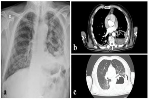
We describe the case of a 67-years-old male patient who was brought in by relatives, with dyspnoea, anorexia and fatique. The patient’s history included a recent diagnosis of a left lower lobe tumor. He was admitted 20 days prior to presentation to a hospital abroad where thoracoscopy was performed for biopsy sampling and potential resection of the lesion. Preoperative medical screening showed good cardiac systolic function with EF 63% and mild lung restriction (FEV1 91%). The procedure was interrupted because the patient suffered heart arrest and was successfully resuscitated. The patient was discharged at his own will, and presented 15 days later febrile (39oC), hypoxic (spO2 84% on air), anuric (SCr 3,97mg/dl), and with significantly elevated inflammatory markers (CRP 582 mg/L, nv<6). Surgical incision was unremarkable. CXR showed inflitrations of the left lower lobe (Figure a). The patient was treated with IV fluids, antibiotics and had two sessions of hemodialysis. Due to clinical worsening he was intubated 36 hours after admission and transferred to the ICU. A CT scan of the thorax was requested which revealed a large left lower lobe abscess and left lung empyema (Figure b,c). He was urgently reoperated, but died a few days later.
Learning point
CT scan is the imaging modality of choice to diagnose post-surgical complications in patients undergoing lung resection.

Figure 1: a. CXR upon admission, b-c CT scan of the thorax
References
- Burel J, Ayoubi ME, Baste J-M, Garnier M, Montagne F, Dacher J-N, Demeyere M. Surgery for lung cancer: postoperative changes and complications-what the Radiologist needs to know. Insights Imaging 2021 Aug 12; 12(1): 116. [DOI: 10.1186/s13244-021-01047-w].
- Motono N, Ishikawa M, Iwai S, Yamagata A, Iijima Y, Uramoto H. Analysis of risk factors for postoperative complications in non-small cell lung cancer: comparison with the Japanese National Clinical Database risk calculator. BMC Surg. 2022 May 14; 22(1): 180. [DOI: 10.1186/s12893-022-01628-6].
- Cardinale L, Priola AM, Priola SM, Boccuzzi F, Dervishi N, Lisi E, Veltri A, Ardissone F. Radiological contribution to the diagnosis of early postoperative complications after lung resection for primary tumor: a revisional study. J Thorac Dis. 2016 Aug; 8(8): E643–E652. [DOI: 10.21037/jtd.2016.07.02].
- Pool KL, Munden RF, Vaporciyan A, O'Sullivan PJ. Radiographic imaging features of thoracic complications after pneumonectomy in oncologic patients. Eur J Radiol 2012 Jan; 81(1): 165-72. [DOI: 10.1016/j.ejrad.2010.08.040].

