Case Report - Volume 2 - Issue 5
Pycnodysostosis: Case report
F Chait*; N Bahlouli; N Allali; S El Haddad; L Chat
Department of Pediatric Radiology, Children’s Hospital, Ibn Sina University Hospital, Rabat, Morocco.
Received Date : Aug 19, 2022
Accepted Date : Oct 10, 2022
Published Date: Oct 26, 2022
Copyright:© Fatima Chait 2022
*Corresponding Author : Fatima Chait, Department of Pediatric Radiology, Children’s Hospital, Ibn Sina University Hospital, Rabat, Morocco
Email: nourrelhoudabahlouli14@gmail.com
DOI: Doi.org/10.55920/2771-019X/1274
Abstract
Pycnodysostosis is a rare genetic osteopathy that combines: dysmorphic syndrome, osteocondensation and growth retardation. This is an newborn of 09 months old in whom a pycnodysostosis was suspected, he was referred by a geneticist to look for radiological signs of this disease. He was born in a first degree consanguineous marriage and had been suffering from repeated spontaneous fractures since birth. The examination revealed a facial dysmorphism with macrocephaly, a persistent anterior fontanel, protruding frontal bumps and micrognathia with a curved aspect of the left lower limb. Skeletal radiographs showed densification of the skull base bones, persistence of the anterior fontanel, hypoplasia of the lower jaw bone and several fractures of different ages with bone calluses, diffuse cortical thickening and metaphyseal enlargement with a short aspect of the upper limb bones were found. In view of the clinical signs and radiological manifestations, the diagnosis retained is pycnodysostosis. Orthopedic management was proposed for the family. Pycnodysostosis is a rare disease, sometimes difficult to diagnose and of delayed onset, which poses a differential diagnostic problem with osteopetrosis and osteogenesis imperfecta.
Introduction
A 09-month-old infant referred by a geneticist to look for radiological signs of pycnodysostosis. From a first degree consanguineous marriage, who presented since birth repeated spontaneous fractures. The examination found Facial dysmorphia made of: Macrocephaly, persistent anterior fontanel, protruding frontal bumps, a prominent nose and Micrognathia, curved aspect of the left lower limb. Skeletal radiographs showed, at the level of the skull: hypoplasia of the lower jaw bone, densification of the base of the skull. In the thorax: hypoplastic aspect of the acromial extremities of the clavicles as well as the ribs several fractures of different ages of the anterior and posterior arches. Limbs: shortening of the upper limb bones, diffuse cortical thickening with a narrow medullary canal and metaphyseal widening, horizontal fracture line with the beginning of a bone callus in the upper metaphysis of the right humerus. There is also a zig-zag fracture line in the lower metaphysis of the left femur with varus displacement. In view of the clinical signs and radiological manifestations, the diagnosis of Pycnodysostosis was retained.
Discussion
Pycnodysostosis, or Toulouse-Lautrec syndrome, is a hereditary condensing osteopathy transmitted in autosomal recessive form. The sex ratio is 1, its exact prevalence is unknown but estimated to be less than 1/100,000, the notion of parental consanguinity is described in 30% of cases [3]. The arguments that allow the diagnosis to be retained are Clinical: characteristic facial dysmorphia (protrusion of the frontal humps, macrocephaly, micrognathia), facial hypoplasia with a prominent nose, spontaneous fractures or fractures caused by a lesser degree of trauma [4]. On standard radiography, the skull is characterized by hypoplasia of the lower jaw, persistence of the anterior fontanel, densification of the base of the skull, and hypoplasia of the acromial end of the clavicle, as in the case of our patient. Fractures of different ages with bone calluses, diffuse osteosclerosis with metaphyseal enlargement and shortening of the limbs [5].
Osteopetrosis remains the main differential diagnosis of pycnodysostosis; it is distinguished by: the normal size, the dense aspect of the bones with disappearance of the medullary canal due to the invasion of the marrow by the bone tissue, which explains the frequency of infectious and hemorrhagic manifestations [2].
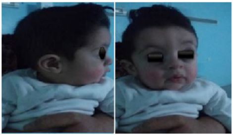
Figure 1: Infant photos showing facial dysmorphia (frontal bumps, micrognathia and macrocephaly).
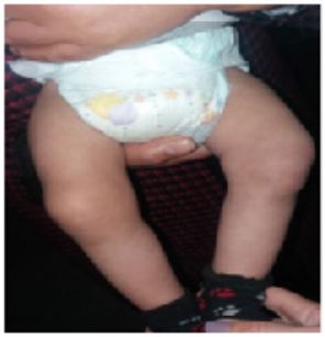
Figure 2: showing the curved aspect of the left lower limb.
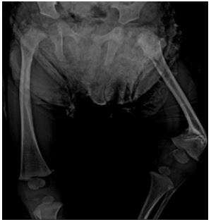
Figure 3: Old zig-zag fracture with varus displacement of the femur, diffuse osteosclerosis, narrow medullary canal.
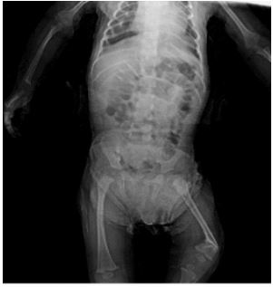
Figure 4: Whole body X-ray showing multiple fractures of different ages of the costal arches, shortening of the limbs.
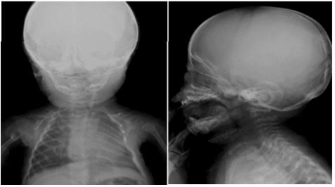
Figure 5: X-ray of the skull (front and side) showing: dental implantation defect, macrocephaly, hypoplasia of the mandible and clavicle, densification of the skull base.
Conclusion
Pycnodysostosis is a rare constitutional bone disease , of sometimes difficult diagnosis, The prognosis is favourable, but the risk of fracture persists[6]. The treatment is essentially preventive.
Conflict of interest: The authors are contributed equally and declare no competing interest.
Contributions: All authors contributed equally in this work.Funding stateme.
Funding statement: The authors received no specific funding for this work.
Ethical Approval: Not required. CONSENT Written consent has been obtained.
Guarantor: Dr. FATIMA CHAIT. Department of Pediatric Radiology, Children’s Hospital, Ibn Sina University Hospital, Rabat, Morocco.
References
- J Nardi, N Meslier. « Pycnodysostose et syndrome d’apnées du sommeil », Médecine Sommeil, 2010; 7(2): 63‑65.
- AE Coudert, P MC. de Vernejoul, « les ostéopétroses Les voies pathogéniques récemment élucidées », Mise Au Point, 2011; 8: 6.
- Q Mujawar, R Naganoor, H Patil, AN Thobbi, S Ukkali, et N Malagi, « Pycnodysostosis with unusual findings: a case report », Cases J, 2(1): 1‑4.
- KKK Ramaiah, GB George, S Padiyath, R Sethuraman, et B Cherian, « Pyknodysostosis: report of a rare case with review of literature », Imaging Sci. Dent., 2011; 41(4): 177-181.
- RJ Bathi et VN Masur, « Pyknodysostosis--a report of two cases with a brief review of the literature », Int. J. Oral Maxillofac. Surg., 2000; 29(6): 439‑442.
- P Maroteaux et M Lamy, « [Pyknodysostosis] », Presse Med., 1962; 70: 999‑1002.

