Clinical Image - Volume 2 - Issue 6
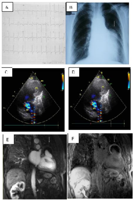
Left Ventricular Aneurism in the Anterobasal Region after Coronary Artery Bypass Surgery
Abdullah Kaplan1* ; Meral Kaplan2
1Department of Cardiology, Kemer Public Hospital, Turkey.
2Department of Radiology, Dinar Public Hospital, Turkey.
Received Date : Sep 16, 2022
Accepted Date : Oct 29, 2022
Published Date: Nov 17, 2022
Copyright:© Abdullah Kaplan 2022
*Corresponding Author : Abdullah Kaplan, Department of Cardiology, Kemer Public Hospital, Yeni Mahalle, Dedeler Mevkii, No 31, Kemer, Antalya, Turkey.
Email: kaplanabd@gmail.com
DOI: Doi.org/10.55920/2771-019X/1296
Abbreviations
LAD: Left anterior descending artery; LIMA: Left internal mammary artery; Cx: Circumflex artery
Clinical Image
A 64 -year-old female was admitted to our inpatient clinic with a 4-month-history of dyspnea and chest pain both at rest and on exertion. One year ago, she had coronary artery by-pass surgery due to triple vessel disease. Electrocardiogram displayed ST segment elevation in leads V2, D1 and aVL (A). A series of cardiac enzyme tests showed no signs of acute myocardial damage. Chest x-ray revealed mild cardiomegaly with a mass image adjacent to the left ventricle (B, arrow). Echocardiogram demonstrated mildly reduced left ventricular function and an aneurysm cavity with a wide neck in the anterobasal region of the left ventricle, partly filled with thrombus (C and D, arrows). Coronary angiography revealed triple vessel disease, total occluded mid LAD, patent LIMA-LAD and safenCx. On a cardiac magnetic resonance imaging (MRI); a left
sided two-chamber cine image clearly showed a thin-walled saccular anterobasal aneurysm partly filled with thrombus (E, arrow), and an inversion-recovery-prepared breath-hold gradient echo cine MRI fifteen minutes after material injection showed delayed enhancement of the wall of the aneurysm and thrombus (F, arrow). There were no coronary vessels surrounding the aneurysm.


