Clinical Image - Volume 2 - Issue 6
A rare cause of chronic respiratory failure in adult
Ana Rita Catarino Ferro* ; Ana Margarida Ferreira Campos
Master of Medicine, Medical Doctor, Pulmonology Department, Centro HospitalarTondela-Viseu, Avenida Rei Dom Duarte, 3504- 509, Viseu, Portugal.
Received Date : Sep 24, 2022
Accepted Date : Oct 29, 2022
Published Date: Nov 18, 2022
Copyright:© Ana Rita Catarino Ferro 2022
*Corresponding Author : Ana Rita Catarino Ferro, Avenida Rei Dom Duarte, 3504-509, Viseu, Portugal.
Email: ana.rita.cf@hotmail.com
DOI: Doi.org/10.55920/2771-019X/1297
Keywords
Congenital heart disease; digital clubbing; respiratory failure.
Clinical Image
Tetralogy of Fallot is the most common form of cyanotic congenital heart disease, characterized by ventricular septal defect, overriding aorta, pulmonary stenosis and right ventricular hypertrophy [1,2].
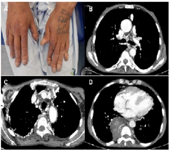
A 61-year-old man, ex-smoker, with previous history of multiple myeloma and tetralogy of Fallot non surgically corrected. Admitted to the Pulmonology Department for worsening dyspnea in the last week. During physical examination, the patient presented peripheral oxygen saturation of 58% and exuberant digital clubbing (Figure 1A). Arterial blood gas analysis revealed respiratory acidemia (pH 7.31, pO2 28, pCO2 49, HCO3- 24.7) and analytical study on peripheral blood showed polyglobulia and thrombocytopenia. No changes on chest radiograph. During hospitalization, he was eupneic, despite maintaining respiratory acidemia with bilevel noninvasive ventilation and never presenting pO2 greater than 40 mmHg with different FiO2 values. Chest CT confirmed the presence of a complex vascular malformation, with a small pulmonary artery diameter and exuberant collateral circulation in the subpleural region of the RUL and left lung fissure (Figure 1 B,C). There was also a right paravertebral mass and some left paravertebral nodules, suggesting extramedullary hematopoiesis (Figure 1D). Transthoracic echocardiogram showed alterations compatible with tetralogy of Fallot with a possible right to left shunt due to interventricular communication and dilated and hypertrophied right ventricle. He was discharged home having a global respiratory failure with FiO2 26% (pH 7.33, pO2 33, pCO2 59, HCO3-31.1) and refused home ventilation.
This case report illustrates a rare cause of chronic respiratory failure in adult, with no relevant clinical improvement with oxygen or noninvasive ventilation therapy. It is also noteworthy for being a patient who refused surgical treatment and has long term survival comparing with other works described in literature [2].
Figure 1: A – Digital clubbing. B, C – Chest CT sowed the presence of a complex vascular malformation with a small pulmonary artery diameter and exuberant collateral circulation in the subpleural region of the RUL and left lung fissure. D – Right paravertebral mass and some left paravertebral nodules, suggesting extramedullary hematopoiesis.
References
- Khan SM, Drury NE, Stickley J, Barron DJ, Brawn WJ, Jones TJ, et al. Tetralogy of Fallot: morphological variations and implications for surgical repair. Eur J Cardiothorac Surg. 2019; 56(1): 101-109. [DOI: 10.1093/ejcts/ezy474].
- Alkashkari W, Al-Husayni F, Almaqati A, AlRahimi J, Albugami S. An Adult Patient with a Tetralogy of Fallot Case. Cureus. 2020; 12(11): e11658. [DOI: 10.7759/cureus.11658].


