Clinical Image - Volume 2 - Issue 6
Imaging findings in pseudohermaphrodite male presenting with complaints of primary infertility
Varsha Mary Khalkho*
Meherbai Tata Memorial Hospital, Dhadkedi, Jamshedpur, Jharkhan 831001, India.
Received Date : Oct 03, 2022
Accepted Date : Nov 15, 2022
Published Date: Dec 08, 2022
Copyright:© Varsha Mary Khalkho 2022
*Corresponding Author : Varsha Mary Khalkho, Meherbai Tata Memorial Hospital, Dhadkedi, Jamshedpur, Jharkhan 831001, India.
Email: varshakhalkho@gmail.com
DOI: Doi.org/10.55920/2771-019X/1316
Clinical Image
A 38 years old male patient came with the history of infertility. His blood reports were normal. He did not have any comorbidities. On ultrasound – there was something behind the urinary bladder which was appearing like uterus. Normal prostate gland was seen in the patient. Patients both kidneys were normal.
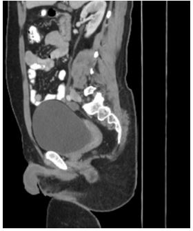
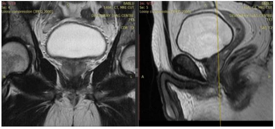
Patients CT scan and MRI was done which showed presence of normal prostate gland. Normal sized uterus and small ovaries were also seen. Uterus measured approximately 6.6 x 2.1cm size. Endometrium was thin and linear.
So, on imaging final diagnosis of male pseudohermaphrodite was made. Teaching point-In cases of infertility patients, it will be useful to carefully image the patient, so the cause of infertility can be ascertained.

Figure 1: Contrast enhanced CT -sagittal image showing uterus (white arrow) and presence of penis.

Figure 2: Non-contrast T2Wt MRI image- coronal image shows normal prostatic tissue (white arrow). Sagittal image shows Presence of uterus adjoining to it presence of prostatic tissue and penis.

