Case Report - Volume 2 - Issue 6
Giant urinary bladder stones, disease of underserved area - The largest case series
Eltahir AE Abdelrahman1; Mosab AA Alzubier2*; Abdolraoof A Abdalla3, Mohammed H Ahmed4
1Surgery Department, University of Alfashir, Faculty of Medicine, Alfashir, Sudan.
2Surgery Department, Gadarif university, Faculty of Medicine, Gadarif, Sudan.
3Urology Department, Sudan Medical specialization Board, Al fashir, Sudan.
4Surgery, State Ministry of Health North Darfur State, Alfashir, Sudan.
Received Date : Oct 18, 2022
Accepted Date : Nov 23, 2022
Published Date: Dec 16, 2022
Copyright:© Mosab AA Alzubier 2022
*Corresponding Author : Mosab AA Alzubier, Surgery Department,Gadarif university, Faculty of Medicine, Gadarif, Sudan.
Email: mosab19103@hotmail.comr
DOI: Doi.org/10.55920/2771-019X/1323
Abstract
Giant urinary bladder stone is defined as a stone weight of more than 100 grams, and more than four centimeters in its longest diameter. It is a rare finding in the current urology practice. To our knowledge, this is the first giant bladder stone series reported in adult Sudanese patients. And it is the longest series worldwide. Here, we report four males with bladder stones (BS) seen in one year. The youngest one was 35 years, and the oldest one was 73 years. Clinical presentation was lower urinary tract symptoms (LUTS) in all cases. One of them was schizophrenic and presented with storage symptoms. Ultrasonography (USS) was done in all cases, and plain x-ray KUB was done in one case. Cystoscopy is done in one case. One of them has obstructing prostate lobes and strawberry stone, he had vesicolithotomy (VL) and transvesical prostatectomy (TVP) simultaneously. All patients underwent open VL. In one of them operation was done under general anesthesia, and three were done under spinal anesthesia. Two of them had vesicocutaeous fistula (VCF), one healed in three months and the other in three weeks. The rest two had an uneventful postoperative course. One of them had a renal impairment that normalized following surgery.
Keywords: Giant bladder stone, bladder stone in Dar Fur, case series, vesicolithotomy, prostatectomy
Introduction
BS accounts for 5% of urinary stone diseases in western countries, and it reaches up to 45% in some African countries [1 – 6]. BS are common in men than in women and children than in adults. [4, 6, 7]. Risk factors that promote BS formation in adults include urinary tract infection (UTI) by urease-producing organisms, chronic urinary retention, and bladder outlet obstruction. These conditions are resultant effects of prostatic diseases, bladder diverticulum, genital prolapse in females, neurogenic bladder dysfunction, urethral stricture, foreign body, and prolonged urethral catheterization [8, 9]. BS is said to be giant when it weighs 100 gm or more and is more than 4 cm in the greatest diameter [3, 5, 6, 10, 11]. The biggest BS weighed 6294 gm and was reported by Arthure in 1953 and found in a bladder diverticulum [4, 6, 10, 12, 13]. The calculus reported by Randall in 1921 weighed 1914 gm and by Powers and Matfferd in 1952 weighed 1410 gm [13]. Charles Azuwike Odoemene reported on three cases of giant BS from Nigeria the largest one weight 1736 gm [8]. Giant BS is a rare condition in developed countries, and fewer than 100 cases were reported worldwide to date.
Case series
Case 1
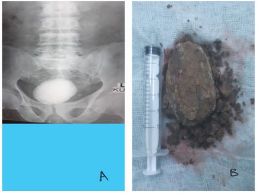
A 73-year-old Sudanese male farmer from north Darfur state presented with burning micturition, urgency, and urgency urinary incontinence for two years. The patient also had a weak stream of urine. He sought medical advice and was treated for UTI all this time. One year ago, the patient passed small stones per urethra spontaneously. On presentation, the patient was unwell and febrile. Digital Rectal Examination (DRE) revealed small prostate with benign clinical features. His lab investigations revealed uncountable pus cells and red blood cells in his urine, serum creatinine of 2.5 mg/dl, and WBC of 18000/HPF. USS showed bilateral hydronephrosis and large BS. X-ray KUB confirmed a big vesical stone, (Fig. 1A). Open VL was done, and a stone of 408 gm was delivered (10 cm in longitudinal diameter). Stone was black, fragile, and very adherent to the bladder. (Fig. 1B). Intraoperative assessment of the prostate was done, and it was not enlarged, bladder neck dilatation was done, and three ways folly's catheter size 22 gauge was fixed. The early postoperative course passed uneventfully. The patient developed VCF that healed three months postoperative. Renal function was normalized. Stone analysis was not done because it is not available.

Figure 1: Case 1 (A, X-Ray (KUB) showing giant bladder stone. B, large bladder stone removed)
Case 2
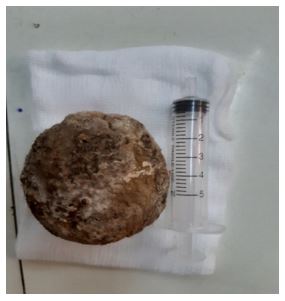
A 41-year-old Sudanese male from north Darfur state presented with burning micturition for one year. On presentation, the patient was unwell and febrile. DRE revealed small prostate. Lab investigations were normal apart from uncountable pus cells and red blood cells in urine and WBC of 10000/HPF. USS showed large BS. Open VL was done, and a stone of 300 gm was delivered (8 cm in longitudinal diameter). Stone was black and hard (Fig. 2). Two ways folly's catheter size 18 gauge was fixed. The early postoperative course passed uneventfully. The patient developed VCF that healed three weeks after surgery.

Figure 2: Large bladder stone removed (case 2)
Case 3
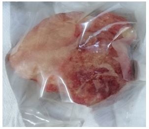
A 36-year-old male presented to the outpatient clinic with increased frequency, urgency, and urgency urinary incontinence noticed by his relatives in the last few months. The patient is schizophrenic and off treatment. Examination revealed an irritable afebrile patient. His lab investigations showed WBC 10000/HPF, urine couldn't be collected for analysis. USS has done with difficulty, and it showed large BS. Open VL was done under general anesthesia. Stone of 300 gm delivered (6 cm in longitudinal diameter). Stone was yellow and hard, (Fig. 3). Two ways folly's catheter size 18 gauge was fixed. The postoperative course passed uneventfully. The patient was referred to the psychiatry department to complete his management.

Figure 3: Large bladder stone removed (case 3)
Case 4
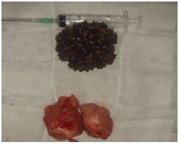
A 65-year-old Sudanese male presented with visible hematuria, burning micturition, urgency, and urgency urinary incontinence for one year. The general examination was unremarkable. DRE revealed a moderately enlarged prostate, with benign features. Lab investigations were within normal, and PSA was 4.2 ng/ml. USS showed a large BS of 6 cm, bilateral multiple renal cysts. Prostate not detected because of large stone shadow that obscures the visualization of prostate. The patient consented to VL and TVP simultaneously. Intraoperative cystoscopy showed normal anterior urethra, enlarged prostate (kissing lateral lobes and small middle lobe) very big BS filling the whole bladder cavity rendering visualization of the entire cavity difficult. Open VL was done, and a stone of 195 gm was delivered (10 cm in longitudinal diameter). Stone was strawberry black. Prostate enucleated (Fig. 4). Three ways folly's catheter size 22 gauge was fixed. Irrigation with normal saline started. The early postoperative course passed uneventfully.

Figure 4: (case 4) Giant bladder stone and prostate were removed surgically.
Discussion
BS is said to be giant when it weighs 100 gm or more and is more than 4 cm in the greatest diameter [3, 5, 6, 10, 11]. The biggest BS weighed 6294 gm and was reported by Arthure in 1953 and found in a bladder diverticulum [4, 6, 10, 12, 13]. The calculus reported by Randall in 1921 weighed 1914 gm and by Powers and Matfferd in 1952 weighed 1410 gm [13]. Charles Azuwike Odoemene reported on three cases of giant BS from Nigeria the largest one weight 1736 gm [8]. Giant BS is a rare condition in developed countries, and fewer than 100 cases were reported worldwide up to date, of them only 30 cases were reported in English literature up to 2018 [9,14,15,16]. Azuwike Odoemene reported three cases in nine years [8].
We see giant BS more frequently as we reported 4 cases in one year. This may be attributed to difficulties to access health facilities in greater Darfur where people need to travel for long distances to reach the optimum health services. Civil wars and conflicts also led people to suffer for reaching optimum health services.
Giant BS formed in non-healthy urinary bladder secondary to bladder outlet obstruction, neurogenic bladder or foreign body, and recurrent UTI [7, 17], in this series one case had obstructing enlarged prostate, one had tough fibrotic bladder neck, the rest two had recurrent UTI of them one is psychotic. BS is suspected in any patient with recurrent UTI not responding to treatment. There was recurrent UTI following urine stasis in all four cases. Urine culture and stone analysis were not done, so the nature of the stones was not specified. In literature, it is postulated that urease-positive organisms are predisposed to magnesium, phosphate, ammonium, and carbonate apatite while E. coli was associated with the formation of calcium oxalate and urate stones [6,8]. Most BS are Struvite (triple phosphate) [4,9,15]. Most patients with giant BS present with storage LUTS including dysuria, increased frequency of micturition, hematuria, suprapubic pain, and/or voiding LUTS as urine retention that may eventually lead to hydronephrosis and renal failure [8]. Only in case one in this series there is bilateral hydronephrosis, hydroureter, and impaired renal function. The delayed presentation may be attributed to post-war internal displacement, poverty, ignorance, and lack of facilities with optimum imaging modalities, this was admitted by other authors in developing countries [8]. Contrast-enhanced CT scan is the modality of choice for diagnosis of urolithiasis including uric acid stones, albeit it is not done for any of our patients in this series [4, 9, and 15]. All patients (100 %) in this series had USS abdomen, it is easy, cost-effective, and available with acceptable accuracy in diagnosing giant BS. One case had plain X-ray KUB. Doumi BA in a comparable geopolitics' area used plain X-ray pelvis or USS to diagnose urinary bladder stones [7]. Cystoscopy was done on one patient to assess the prostate and to visualize the bladder wall for other pathology as bladder tumors may coincide with BS [12]. In this series, cystoscopy was useful in the assessment of the prostate, but it was difficult to visualize the entire bladder wall because the stone filled almost the whole cavity. All cases in this series underwent open VL. Three of them were under spinal anesthesia and one was done under general anesthesia because of his psychiatric illness. Open VL is the modality of choice for the treatment of giant BS, it is easy to perform, no need for a drain or catheter unless the procedure is accompanied by other procedures like prostatectomy done simultaneously. [4, 9, 12, 15, 18]. Endoscopic procedures though it has advantages in evaluating and correcting urethra and bladder neck obstruction, it is not suitable for such large stones. [9, 15]. Tan J, Singh reported on the spontaneous passage of two giant bladder stones in females [14].
Conclusion
To the best of our knowledge, this is the largest case series of giant BS worldwide. Though giant BS is becoming rare in the modern urology practice in developed countries, we saw four cases in one year. Lack of optimum health services, ignorance, and illiteracy all are contributed to the delayed presentation of patients with BS in this series.
List of abbreviations
BS; bladder stone. LUTS; lower urinary tract symptoms. VL; vesicolithotomy. TVP; transvesical prostatectomy. USS; ultrasound. VCF; vesicocutaneous fistula. DRE; digital rectal examination. UTI; urinary tract infections.
References
- Mbonu, O., Attah, C. & Ikeakor, I. Urolithiasis in an African population. International Urology and Nephrology 16, 291–296 (1984). https://doi.org/10.1007/BF02081863
- Rahman GA, Akande AA, Mamudu NA (2005) Giant vesical calculi: Experience with management of two Nigerians. Nig J Surg Res 7:203–205. https://doi.org/10.4314/njsr.v7i1.12280
- LawalA T, Ahmed M, Bello A, Maitama H Y, Awaisu M (2016) Atypical presentation of enormous vesical calculus: A case report. Afr J Urol; 22:207–209. http://dx.doi.org/10.1016/j.afju.2015.07.005
- Garg, V., Awasthi, R. C., Jain, G., Pandey, S., & Maheshwari, M. K. (2016). Giant Vesical Calculus. International Journal of Medical and Dental Sciences, 1265–1268. https://doi.org/10.19056/ijmdsjssmes/2016/v5i2/100618
- Schwartz BF, Stoller ML. The vesical calculus. Urol Clin North Am. 2000 May;27(2):333-46. doi: 10.1016/s0094-0143(05)70262-7. PMID: 10778475.
- Diniz, A.L.L., Vieiralves, R.R., de Souza, T.A. and Favorito, L.A. (2017) Giant Bladder Stone and Renal Failure: A Case Report, Literature Review and Future Perspectives. Open Access Library Journal, 4: e3332. http://dx.doi.org/10.4236/oalib.1103332
- Doumi BA (2008) Vesical calculi in El Oeid Hospital: Sudan JMS 3: 1. https://pdfs.semanticscholar.org/ad1f/83bd32765b186affda28d97fefb4eb76bfb6.pdf
- Odoemene CA. Giant urinary bladder stones. Int J Case Rep Images 2018; 9:100933Z01CO2018. doi: 10.5348/100933Z01CO2018CS
- Shilpi G, Onkar S, Sumit S, Mathur Raj K (2017 Giant vesical calculus. The Internet Journal of Surgery;17:(1). https://ispub.com/IJS/17/1/10317#
- ARTHURE H. A large abdominal calculus. J Obstet Gynaecol Br Emp. 1953 Jun;60(3):416. doi: 10.1111/j.1471-0528. 1953.tb14080. x. PMID: 13062008.
- Trinchieri A. Epidemiology of urolithiasis: an update. Clin Cases Miner Bone Metab. 2008 May;5(2):101-6. PMID: 22460989; PMCID: PMC2781200. http://www.ncbi.nlm.nih.gov/pmc/articles/pmc2781200/
- Kirakoya B, Kabore M, Pare AK, Abubakar BM. GIANT BLADDER STONE IN A PATIENT WITH TUMOR OF THE BLADDER: A RARE CO-MORBIDITY. J West Afr Coll Surg. 2018 Jul-Sep;8(3):114-120. PMID: 32754460; PMCID: PMC7368580. http://www.ncbi.nlm.nih.gov/pmc/articles/pmc7368580/
- Butt MK, Inam A, Rehman S (2015) Giant vesical stone. A case reports. APMC 9:48–51. https://apmcfmu.com/index.php/apmc/article/download/345/283/
- TAN, J., SINGH, P., HUANG, E., HAN, H.. Spontaneous Expulsion of Giant Bladder Stone: Case Report. Journal of Medical Cases, North America, 9, jan. 2018. Available at: <https://www.journalmc.org/index.php/JMC/article/view/2850/2320>
- Mylarappa P, Amey P, Prathvi, Sandeep P, Banale KB, Ramesh D(2012) Giant pelvic cocoon with intra prostatic extension: Case report. Asian Journal of Medical Science 3:47–49. http://dx.doi.org/10.3126/ajms.v3i4.7160
- Braja Mohan Mishra, Malay Krushna Nayak, Rabinarayan Guru, Sandip Mishra. “A Rare Giant Urinary Bladder Stone in an Elderly Male: A Case Report”. Journal of Evolution of Medical and Dental Sciences 2015; Vol. 4, Issue 20, March 09; Page: 3558-3561, DOI: 10.14260/jemds/2015/513
- Faisal Ahmed et al. Giant urinary bladder calculi in a 60-year-old man: a case report. Pan African Medical Journal. 2022; 41:78. [doi: 10.11604/pamj.2022.41.78.33131]. https://www.panafrican-med-journal.com/content/article/41/78/full
- Makkawi MAE, Babikir WIE, Elhag MM (2019) Giant Urinary Bladder Stone Formed Around a Pin in a Young Sudanese Girl: A Case Report and Literature Review. Clin Surg 4: 24-36. http://www.clinicsinsurgery.com/open-access/giant-urinary-bladder-stone-formed-around-a-pin-in-a-young-sudanese-girl-a-case-report-and-literature-review-3849.pdf

