Research Article - Volume 2 - Issue 6
A rare anatomical variation of the hand lumbrical muscles: A case report and literature review
Alireza Shams1*; Ali Ramadan2; Mohmmadamin Shams3
1Department of Anatomy, Faculty of Medicine, Alborz University of Medical Sciences, Karaj, Iran.
2Faculty of Medicine, Alborz University of Medical Sciences, Karaj, Iran.
3Faculty of Medicine, Iran University of Medical Sciences, Tehran, Iran.
Received Date : Oct 31, 2022
Accepted Date : Nov 29, 2022
Published Date: Dec 21, 2022
Copyright:© Alireza Shams 2022
*Corresponding Author : Alireza Shams, Department of Anatomy, Faculty of Medicine, Alborz University of Medical Sciences, Karaj, Iran.Tel: +989123900319 Fax: + 982634287311
Email: dr.shams@abzums.ac.ir
DOI: Doi.org/10.55920/2771-019X/1327
Abstract
Background/Objective: We report a case of unusual anatomical variation and rare abnormality of the lumbrical muscles (LM) in a cadaver after dissection. Usually, the hand LM arises from the flexor digitorum profundus (FDP) tendons and inserts into the extensor hoods of the corresponding fingers. LM is fundamental in the performance of fine skillful movements of the hand.
Patient Report: During routine dissection on a cadaver donated for medical education, we observed unusual origin and morphology of both first LM of a male cadaver. We followed Cunningham's dissection manual as a guide. We observed that the 1th LM in both hands had an unusual origin from the first tendon of Flexor Digitorum Superficialis (FDS) instead of FDP.
Conclusion: Most muscles begin and insert onto bones, but the LMs originate and insert onto tendons. Due to LM originating from the palmar aspect of the hand and their insertion onto the extensor expansion of the dorsal aspect of digits, Deformities of intrinsic muscles of the hand can cause an extensive range of disabilities in movements. Surgeons must be aware of these variations to keep LMs safe and avoided. Operations like flexor tendon repair, median nerve release, and deep laceration repair using stitch are straightly focused on this region. Due to their proximal extension, it might lead to carpal tunnel syndrome. Furthermore, the anomalous origin, length, and volume of LM are essential parameters for the outcome of operations on the carpal tunnel.
Keywords: Variation; Lumbrical Muscles; Gross anatomy; Dissection; Hand operation.
Introduction
The lumbrical muscles are named after the Latin word "lumbricidae," meaning earthworm. This is due to their unique appearance. Usually, there are four lumbrical muscles in a human hand. All originate from FDP tendons and insert onto the extensor expansions on the lateral side of the corresponding digits. The lumbricals are unique because although most muscles originate and insert onto bones, LMs both originate and insert onto tendons, giving the lumbricals moveable attachments. These muscles participate in metacarpophalangeal joint flexion and interphalangeal joint extension. Complex digital movements of the human hand have been described as a revolution in evolution. This distinctive feature makes determining the function of this muscle very difficult because the utility of the lumbricals depends upon the activity of the common extensor tendon and the position of the finger joints [1]. LMs, as a significant component of the intrinsic part of hand musculature, display complex actions, thus making it possible for the human hand to be precise and dexterous. Many authors have reported variations of the LMs. However, the variations of the first and second lumbrical muscles seem to be rarer than those of the third and the fourth [2]. An example would be the case report of Mehta et al. about the anomalous originof the first lumbrical muscle [3]. Anomalies in the morphology of the LMs are uncommon, but studying them is necessary to maximize the success rates of hand surgical operations.
Accessory first lumbrical muscle bellies have been found arising from the deep layer of FDS [4] and FDP [5] in the forearm. Furthermore, there are reports of an accessory first lumbrical muscle belly arising from the radial side of the FDP tendon near the proximal border of the transverse carpal ligament [6].There have also been reports of a tripennate first lumbrical muscle originating from three unique sources [7], suggesting that the first lumbrical muscle demonstrates different variations. Abnormalities of the intrinsic muscles of the hand can lead to a wide range of inabilities in movements.These variations can also increase the risk of CTS [8,9]. The first and second lumbricalsarise from the radial side of the FDP tendons of the second and third fingers. Thus, the first and second lumbricalsare unipennate, whereas the third and fourth are bipennate. Two ulnar and two radial lumbrical muscles are innervated by the ulnar and median nerves, respectively [10].
We reported a rare bilateral variation of the first lumbrical muscle, discovered incidentally in gross anatomy dissection class. In the present case, we observed that the first lumbrical muscle in both hands took origin from the FDS tendon, and the clinical and surgical significance of the present case was discussed.
Case presentation
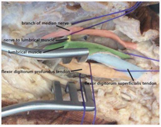
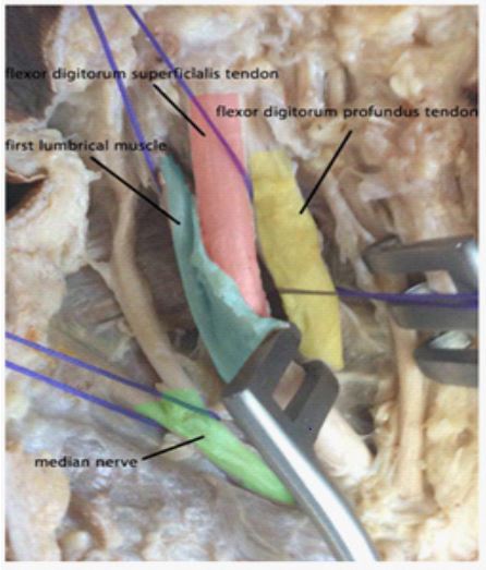
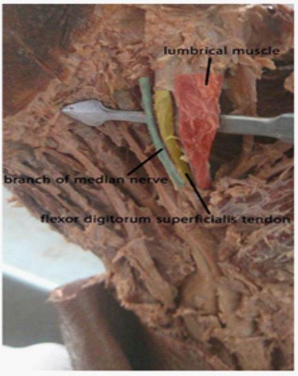
During routine dissection of the teaching program of medical students in the anatomy learning center of the medicine faculty, unusual origin and morphology of lumbrical muscles in both hands of a 50-year-old male cadaver were observed [Figure 1]. We followed Cunningham's manual of dissection as a guide. After incising the skin and subcutaneous tissue, we identified lumbricals, specifically the first lumbrical, by incising the thinner space located deep to the flexor tendinous sheath [Figure 2, 3]. We dissectedthe palmar aponeurosis inferiorly using scalpel No. 11 and forceps for better exposure. The lumbrical tendon was transmitted distally anterior to the deep transverse metacarpal ligament and inserted normally to the lateral side of the expansion hood for the index finger as expected. Usually, the four lumbrical muscles originate from the tendons of FDP and insert onto the extensor expansions on the radial side of the corresponding fingers [11].
In this specific case, we discovered that the first lumbrical originates from the lateral side of FDS instead of FDP. (70mm in length and 7 mm in width) a twig from the median nerve innervated both of the first lumbrical muscles in the right and left hand [Figure 4]. We also found another anomaly in the upper limb. The musculocutaneous nerve had a common trunk with a lateral branch origin of the median nerve instead of the lateral cord of the brachial plexus.

Figure1: Dissection of the right hand of a male cadaver, first Lubmrical (Red-colored) originates from Flexor digitorumsuperficialis muscle tendon (Blue colored) instead of Flexor DigitorumProfundus muscle tendon (Orange colored). The branch of the Median nerve into the first Lumbrical was exposed (Yellow colored). Note that the nerve and attachment of the lumbrical indicate the VA variation of the origin of the first lumbrical.

Figure 2: Dissection of the right hand, first lumbrical muscle (blue-colored) originates from the flexor digitorumsuperficialis tendon (red-colored). Deep to the flexor digitorumsuperficialis, the flexor digitorumprofundus tendon (yellow-colored) is visible.

Figure 3: Dissection of the left hand, the first lumbrical (red-colored) originates from the flexor digitorumsuperficialis tendon (yellow-colored). The common palmar branch of the median nerve (blue-colored) can also be seen.

Figure 4: Dissection of the left hand, the image shows a complete dissection of the first lumbrical muscle(red-colored). The flexor digitorumsuperficialis (green-colored) and flexor digitorumprofundus (blue-colored)tendons are also shown here.
Discussion
The lumbricals flex the metacarpophalangeal joints and extend the interphalangeal joints. These muscles have more clinical relevance than the lumbricals of the foot because of their proximity to the carpal tunnel and their role in the precision of the hand. The abnormality in these muscles is of great clinical importance.Even though the third lumbrical is considered the most variant and the fourth lumbrical the most absent [3,12], the morphological variations of the first and second lumbricals should not be overlooked.Butler et al. reported a case where a tendon arising in the index lumbrical was inserted proximally into the FDS of the index [13]. Soubhagya et al. also reported an additional muscle belly of the first lumbrical muscle that took origin from the FDS tendon to the second finger close to the proximal margin of the flexor retinaculum [14]. Furthermore, in the present case, we observed that the abnormal first lumbrical muscles took origin from the FDS tendons instead of their usual origin.It should also be considered that since the anomaly is seen in both limbs, this variation could have genetic origins. However, we could not further investigate the genetic origins of this case since we did not have access to the subject's family's medical records.
The lumbrical muscles play a vital role in the complex and precise movements of the human hand; thus, any variation in these muscles could limit the range of motion and lower strength and cause different complications for patients. The lumbrical muscles are thought to be the principal cause of camptodactyly. In a series of 74 consecutive operations for camptodactyly, McFarlane et al. discovered that the fourth lumbrical muscle to the small finger had an anomalous insertion in all cases [15]. Variations of lumbrical have been reported as a culpritcausing carpal tunnel syndrome (CTS) [16]. Any proximal attachment of LMs extending into the carpal tunnel may limit the carpal space, thus compressing the median nerve, leading to CTS. Many studies have reported a higher origin of the first and second lumbricals in the carpal tunnel. For example, Benny et al. reported an army veteran with CTS resulting from an abnormal origin of the LMs proximal to the carpal tunnel in the forearm arising from both FDS and FDP tendons. He had found a hypertrophic muscle compressing the median nerve [17]. Silawal et al. described a first left lumbrical with two muscle bellies, one originating from the medial condyle of the humorous and one originating from the first tendon of the FDP; the belly originating from the humerus hadentered the distal part of the carpal tunnel [18]. A similar observation was reported in a study by Singh et al. showing a bipennate origin of the 1st lumbrical, extending from the distal part of the forearm [5]. In another study done by Eriksen, a 31-year-old male worker suffered from CTS and had a lack of strength in his right hand due to the abnormally proximal origin of the third lumbrical muscle. In this case, the compression of the median nerve only occurred after exercise, and there was no pressure on the median nerve during rest [19].
There are also some reports of absence in LMs. In a study by Priyadharshini and Kumar, the third and fourth lumbricals were absent in both hands of a 55 years old male cadaver [20]. Baa et al. reported aunilateral absence of the second, third, and fourth lumbrical muscles [21]. Of 30 cadavers examined in a study done by Hosapatna M et al., the third lumbrical was absent in one case (3.3%) [22]. Clinicians and Surgeons should be aware of these variations while designing and dealing with hand surgeries. Operations like flexor tendon repair, digital branches from median nerve release, and deep laceration repair using stitch are straightly focused on this region. An attempt has been made to understand its clinical, embryological, and phylogenetic aspects. A hand surgeon might think there are no vital structures around the FDS tendon and cut any tissue he sees blindly. However, as is mentioned, lumbricals may have origins in the FDS tendon, and neglecting these variations could have consequences for patients.
Conclusions
Surgeons must be aware of these variations while dealing with the hand in order to keep muscles safe and avoided during surgical procedures.The anomalous variation of these lumbricals may lead to confusion during surgical operations, which could lower the success rate of these medical procedures. Therefore the more knowledge we have about these variations, the more chance we can successfully perform operations in this region.
Conflicts of interest and source of funding: None declared.
Acknowledgments
The authors wish to thank the individuals who donated their bodies to science and the progress of medicine. The data from such research can help improve the result of surgical operations and expand humankind's overall knowledge of the human body. On that account, these donors and their families deserve our highest regard and gratitude.
References
- Singh S, Loh HK, Mehta V. Variant lumbrical musculature of the left hand: Clinico-anatomic elucidation. Morphologie. 2016; 100(331): 256-259.
- Parminder K. Morphological study of lumbricals - a cadaveric study. J ClinDiagn Res. 2013; 7(8): 1558-60.
- Mehta HJ, Gardner WU. A study of lumbrical muscles in the human hand. Am J Anat. 1961; 109: 227 38.
- Koizumi M, Kawai K, Honma S, Kodama K. Anomalous lumbrical muscles arising from the deep surface of flexor digitorumsuperficialis muscles in man. Ann Anat. 2002; 184: 387-392.
- Singh G, Bay BH, Yip GW, Tay S. Lumbrical muscle with an additional origin in the forearm. ANZ J Surg. 2001; 71: 301-302.
- Nayak SR, Rathan R, Chauhan R, Krishnamurthy A, Prabhu LV. An additional muscle belly of the first lumbrical muscle. Cases J. 2008; 1: 103.
- Nation HL, Jeong SY, Jeong SW, Occhialini AP. Anomalous muscles and nerves in the hand of a 94-year-old cadaver—A case report. Int J Surg Case Rep. 2019; 65: 119-123.
- Zhang G, FendersonBA. Bilateral accessory flexor muscle of the forearm giving rise to a variant head of the first lumbrical. Int J Anat Variations. 2015; 8: 4-6.
- De Smet L. Median and ulnar nerve compression at the wrist caused by anomalous muscles. Acta Orthop Belg. 2002; 68: 431-8.
- Burlakoti A, Massy-Westropp N, Wechalekar H. Variant third and fourth lumbrical muscles of the left hand.Int J AnatVar (IJAV). 2013; 6: 93-95
- Windisch G. The fourth lumbrical muscle in the hand: a variation of insertion on the fifth finger. SurgRadiol Anat. 2000; 22(3-4): 213-5.
- Koizumi M, Kawai K, Honma S, Kodama K. Anomalous lumbrical muscles arising from the deep surface of Flexor digitorumsuperficialis in man. Ann Anat. 2002; 184: 387 92.
- Butler B Jr, Bigley EC. Aberrant index (first) lumbrical tendinous origin associated with carpal tunnel syndrome. J Bone Joint Surg [Am]. 1971; 53: 160-2.
- Nayak SR, Rathan R, Chauhan R, Krishnamurthy A, Prabhu LV. An additional muscle belly of the first lumbrical muscle. Cases J. 2008; 1(1): 103.
- McFarlane RM, Classen DA, Porte AM, Botz JS. The anatomy and treatment of camptodactyly of the small finger. J Hand Surg Am.1992; 17(1): 35-44.
- Potu BK, Gorantla VR, Rao MS, Bhat KM, Vollala VR, Pulakunta T, Nayak SR. Anomalous origin of the lumbrical muscles: a study on South Indian cadavers. Morphologie. 2008; 92(297): 87-9.
- Benny E, Parminder GS, NurAzuatul AK, Tan JA, Ahmad Suparno B, et al. Anatomical Variations of the Lumbrical Muscles Causing Carpal Tunnel Syndrome. Med and Health. 2019; 14(1): 197-202.
- Silawal S, Galal KR, Schulze-Tanzil G. A rare variation of intrinsic and extrinsic hand muscles represented by a bi-ventered first lumbrical extending into the carpal tunnel combined with bilateral fifth superficial flexor digitorum tendon regression. Morphologie. 2018; 102(339): 294-301.
- Eriksen J. A case of carpal tunnel syndrome on the basis of an abnormally long lumbrical muscle. ActaOrthop Scand. 1973; 44(3): 275-7.
- Priyadharshini NA, Kumar VD, Rajprasath R. Bilateral absence of third and fourth lumbricals: A case report with clinico-evolutionary insight. J Curr Res Sci Med. 2018; 4: 116-8.
- Baa J, Prusti JS, Rath S. Unilateral absence of second, third and fourth lumbricals: A rare case report with an evolutionary significance. Int J Anat Res. 2015; 3: 1237 9.
- Hosapatna M, Bangera H, Kumar N, Sumalatha S, Nitya. Morphological variations in lumbricals of hand – A cadaveric study. Int J. 2013; 2013: 821692.

