Clinical Image - Volume 3 - Issue 1
Dermoscopy of vulvar bowen disease
Nada Bennouna1*; Kenza Baline2; Fouzia Hali2; Soumiya Chiheb2
1Department of Dermatology and Venerology, Ibn Rochd University Hospital Casablanca, Morocco.
2Department of Dermatology and Venerology, Ibn Rochd University Hospital, 1 rue des Hôpitaux -ex Banaflous, 20360 Casablanca Morocco.
Received Date : Nov 11, 2022
Accepted Date : Dec 15, 2022
Published Date: Jan 05, 2023
Copyright:© Nada Bennouna 2023
*Corresponding Author : Nada Bennouna, Department of Dermatology and Venerology, Ibn Rochd University Hospital Casablanca, Morocco.
Email: nadabennouna1@gmail.com
DOI: Doi.org/10.55920/2771-019X/1340
Clinical Image
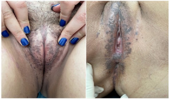
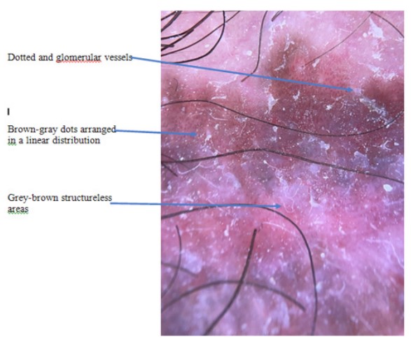
A 26-year-old-women, with a history of cutaneous Rosai Dorfman disease treated with oral corticosteroids and methotrexate, presented with a 5 months history of multiple gray-brown papules and small well-defined patches located symmetrically on her vulval labia and around the anus (Figure 1). Genital warts, a verrucous nevus and Bowen disease were suspected among our clinical diagnosis. The dermoscopy revealed a pigmented papillomatous surface, brown-gray dots arranged in a linear distribution, grey-brown structureless areas and widespread dotted and glomerular vessels (Figure 2). A punch biopsy was done, and the histology showed highly atypical squamous cells accompanied by cytological atypia and mitosis with multiple koilocyte, consistent with the diagnosis of bowen disease. The patient was treated with topical 5-fluorouracil once daily for four weeks.
Keywords: Bowen disease; Vulvar; Dermoscopy.

Figure1: multiplegray-brown papules and small well-defined patches located symmetrically on her vulval labia and around the anus.

Figure2: dermoscopy image.
Acknowledgements: None.
Source of Support: None.
Conflict of Interest: The authors declares that there is no conflict of interest.

