Clinical Image - Volume 3 - Issue 1
Vestibular schwannoma: Ice cream cone sign
Berrada Kenza*; El Harass Yahya; Amalik Sanaa; Latib Rachida; Omor Youssef
National Institute of Oncology, Mohammed V University in Rabat, Rabat, Morocco.
Received Date : Nov 17, 2022
Accepted Date : Dec 27, 2022
Published Date: Jan 17, 2023
Copyright:© Berrada Kenza 2023
*Corresponding Author : Berrada Kenza, Radiology Department, National Institute of Oncology, Mohammed V University in Rabat, Rabat, Morocco.
Email: Knouz.berrada@gmail.com
DOI: Doi.org/10.55920/2771-019X/1350
Abstract
Vestibular schwannoms is a benign extra-axial tumor the most frequent cerebellopontine angle masses. It can interfere with the face nerves, causing facial numbness and paralysis. MRI is the most specific and first intention examination to make the diagnosis.
Keywords: Vestibular schwannoma- MRI- Ice cream cone sign.
Vestibular schwannomas also called acoustic neuromas, are extra-axial slow-growing benigntumors developing from Schwann cells of the vestibular portion of the 8th cranial nerve. These tumors account for ~8% of all primary intracranial tumors .They are due to an overproduction of Schwann cells .The tumor, which develops in a closed and rigid environment (bone, skull), will compress the nerve fibers intended for the auditory systems usually causing unilateral or asymmetric hearing loss. As the tumor grows, it can interfere with the face nerves (the trigeminal and facial nerves), causing facial numbness and paralysis.
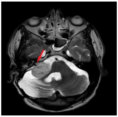
Bilateral vestibular schwannomas is mostly associated to the presence of neurofibromatosis type 2 (NF2). This genetic disease is characterized by the development of meningiomas, spinal cord tumors, and schwannomas in multiple cranial nerves. Radiologically, T1-weighted MRI sections demonstrate the presence of an expansive mass of the left pontocerebellar angle that extends into the ear canal. This mass is slightly more hypointense than the cerebellar parenchyma. It compresses the side edge of the pons. It has an ice cream cone appearance. The intracanalicular component represents the cone and the cerebellopontine angle (CPA) (cisternal) component representing the ice cream ball (Figure 1).

Figure 1: Axial section MRI weight T2 showing the ice cream cone sign on the right (arrow).

