Case Report - Volume 3 - Issue 1
A 4-year-old girl’s radiological finding of hyperparathyroidism as a result of chronic renal failure
Abourak Chaimae*; Oukassem Siham; El Houssni Jihane; Salmane Mehdi; Chat Latifa; Allali Nazik; El Haddad Siham
Department of radiology, Children’s hospital, UHC Ibn Sina, Mohamed V University, Rabat, Morocco.
Received Date : Nov 25, 2022
Accepted Date : Jan 02, 2023
Published Date: Jan 23, 2023
Copyright:© Abourak Chaimae 2023
*Corresponding Author : Abourak Chaimae, Department of radiology, Children’s hospital, UHC Ibn Sina, Mohamed V University, Rabat, Morocco.
Email: chaymaabourak@gmail.com
DOI: Doi.org/10.55920/2771-019X/1355
Abstract
The most frequent bone abnormalities in people with chronic renal failure are caused by hyperparathyroidism. On a clinical level, it is caused by a variety of reasons and has a protracted clinical asymptomatic period. We described the case of a 4-year-old girl with torticollis, a hyper cervical lesion, and a scoliotic attitude who was being monitored for chronic renal failure. A radio graphic check-up was ordered in this instance to look for bone indications of hyperparathyroidism owing to chronic renal failure. The primary radiographic bone indicators of hyperparathyroidism owing to chronic renal failure are described in this article, and they are numerous, varied, and crucial for an efficient surveillance of this population of disorders.
Keywords: Chronic renal insufissance; secondary parathyroidism; osteoadystrophy; osteolytic lesions ; osteosclerosis
Introduction
Early phospho calcicum metabolism difficulties brought on by chronic kidney failure gradually lead to secondary hyperparathyroidism, which is accompanied by other disorders such acidosis, nutritional issues, loss of mobility, and chronic inflammation. According to the severity of kidney failure, each of these factors has negative effects on the bone to varied degrees [2].
Case Report
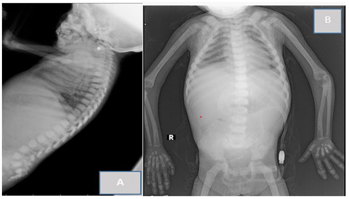
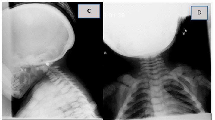
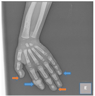
A 4-year-old girl on peritoneal dialysis for chronic renal failure has a torticollis with cervical hyper-flexion and scoliosis, dorso-lumbarbone condensation in the cervical spine, as well as medullary enlargement in both clavicles (Figures A, B, C, and D). An X-ray of the left hand also shows stunting and osteolysis in the joints of the first, third, and fourth fingers, as well as All of these indicators point to secondary hyperparathyroidism, which was in fact validated by the biological evaluation.

Figure A and B: Standard radiography of the cervico dorso lumbarspine, A: profile, B: face, objectifying diffuse bone condensation.

Figure C and D: Radiography of the cervical spine, C profile; D: face. Showing bilateral clavicular medullary enlargement

Figure E: X-ray of the left hand, Estimated boneage of 2 years according to the Greulich and Pyle method, osteolysis of the houppes of the distal phalanx of the 1st ,3eme and 4th fingers bone outgrowth next to the distal phalanx of the 5th finger, of the middle and distal phalanx of the 2nd finger with pinching of the inter-phalangian articular space next to it, diffuse bone condensation
Description
The most frequent bone abnormalities in chronic renal failure are brought on by hyperparathyroidism. It begins early, and its severity is inversely correlated with the length of renal failure. It is caused by a number of reasons, the oldest of which is phosphate retention associated with the progressively declining number of functioning nephrons. As a result of parathyroid gland hyperplasia and increased bone secretion of FGF 23, it is characterized by a higher amount of circulating parathormone. FGF23 functions as parathormone (PTH), which is why phosphate excretion through the urine is increased. The level of bon eremodeling is increased by secondary hyperparathyroidism, which causes fibrous osteitis lesions by stimulating osteoblastic production and osteoclastic resorption [1].
Osteodystrophy is a long-lasting asymptomatic condition that can emerge clinically as growth retardation, bone discomfort, pathological fractures, and, in more severe cases, drumstick-like fingers due to the destruction of distal phalanges, tendon ruptures, and, most notably, calciphylaxia [2]. From a radiological perspective, hyperparathyroidism results in condensant osteolesions, diffuse osteopenia, localized osteolytic lesions, and it plays a role in the development of periosteal appositions. It is possible to see soft tissue calcifications and articular juxta erosions. The sizes of localized osteolytic lesions range from microgaps to macrogaps, geodes to cystic cavities. Different forms of resorption are identified based on their topographies: periosteal, intracortical, (endostated), chondral, ligamentary, and trabecular.
Both adults and kids are notably affected in the hands. One of the earliest radiological symptoms of hyperparathyroidism is reaching Phalangian houppes. Their cortex vanishes, then tiny scratches in the shape of a "postage stamp" appear. The involvement of which would be the radiological sign best connected to histological abnormalities, subperiosteal erosions can also damage the diaphysis of the phalanges, particularly the radial edge of the second and third phalanges. The cortical's grooves and irregular surfaces are caused by subperiosteal and endostal resorption.
When bone degradation is advanced, corticales become extremely thin and it can be challenging to distinguish between individual pictures. In extreme cases, a major resorption of the phalangette's diaphys is might result in band osteolysis of the third phalange's middle portion. Radiologically, osteosclerosis is characterized by an increase in bone density, and histologically, it is characterized by an increase in total bone tissue, including both osteoid and mineralized tissue. As a result of the bottom and higher plateaus of many vertebrae condensing, it frequently occurs at the spinal level and gives the appearance of sandwich vertebrae. It is especially common during renal failure [1]. Osteoporosis is the main condition that needs to beruled out. The major goal of treatment for children with secondary hyperparathyroidism is to maintain a stable PTH level that corresponds to a normal rate of osseous remodeling that will support these young patients' growth [2].
Conclusion
The majority of kids on dialysis for renal insufficiency have phosphocalcic metabolic issues that frequently result in secondary hyperparathyroidism, which is the cause of bone deformities. His radiological semiology is comprehensive and diverse. A thorough understanding of the pathophysiology of the lesions seen is necessary for successful radiological monitoring of chronic hemodialysis, as is an interpretation of the pictures based on clinical-biological correlations [1,2].
Competing Interests: The authors declare that they have no links of interest .
Informed Consent: Patient consent has been given.
Conclusion
Sclerosing mesenteritis is a rare presentation of IgG4-RD. It may present as a mass which may lead to a misdiagnosis of malignancy. The gold standard for diagnosis is via tissue specimen and histopathological diagnosis. Treatment may involve surgical excision and treatment with steroid or steroid-sparing agents. If refractory, can be treated with rituximab. Early diagnosis and treatment are important to avoid fibrosis.
Declaration of interest: None.
Funding: None.
Acknowledgments: None.
References
- B Grignon, O Regent, P Pere, M Kessler, P Nelter, A Gaucher. Osteoarticular manifestations in chronic hemodialysis patients. Current aspects. Radiology sheets. Mason. Paris. 1987; 27(n° 6): 411-425.
- Hinchi H. Hyperparathyroidism in children on chronic dialysis: about 13 cases. Medicine thesis Rabat. 2021; 213.

