Clinical Image - Volume 3 - Issue 1
An unusual occurrence of abdominal neurofibroma with the neck and orbit plexiform neurofibroma in the pediatric age group
Mohammed J Aboud1*; Shaimaa M Kadhim2; Noor M Abudi3; Haidar M Joudi4; Zeena M Joudi4
1Pediatric surgery unit, the Maternity and Child Teaching Hospital, Al Diwaniya, Al Qadisiya, Iraq.
2Department of pediatrics, the Maternity and Child Teaching Hospital, Al Diwaniya, Al Qadisiya, Iraq.
3Department of surgery, Al Diwaniya General Teaching Hospital, Al Qadisiya, Iraq.
4Graduated doctor, College of Medicine, Al Qadisiya University, Iraq.
Received Date : Dec 13, 2022
Accepted Date : Jan 07, 2023
Published Date: Jan 29, 2023
Copyright:© Mohammed J Aboud 2023
*Corresponding Author : Mohammed J Aboud, Pediatric surgery unit, the Maternity and Child Teaching Hospital, Al Diwaniya, Al Qadisiya, Iraq.
Email: mohammedabud@yahoo.com
DOI: Doi.org/10.55920/2771-019X/1360
Clinical Image
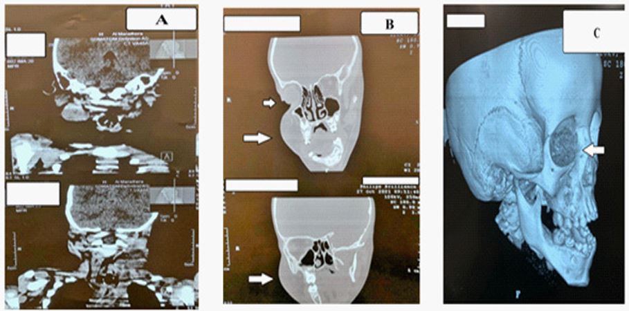
Peripheral nerve sheath tumors, specifically neurofibromas and schwannoma variants, occur both sporadically and in the setting of hereditary conditions, including neurofibromatosis type 1 (NF1), neurofibromatosis type 2 (NF2), and familial/sporadic schwannoma [1]. It is a frequent and polymorphic genetic disorder. The severity is related to the complications. The degeneration of neurofibroma is a very rare complication of neurofibromatosis [1]. Neurofibromatosis type 1 is a multisystemic disorder that can affect several organs [2]. Sporadic gastrointestinal neurofibroma occurs considerably less often [3]. It may have a course of abdominal pain, discomfort, nausea, dyspepsia, and vomiting. Many of these tumors are age-related, occur at specific anatomic locations, and have unique imaging features [2,3]. In literature, many patients have a variety of organs affected because there is a high prevalence of multiple tumors occurring in the same patient [3]. A 5-year-old male patient was admitted to our pediatric surgery unit with the complaint of abdominal pain and distention. The family noticed the development of pigmented patches at birth on his skin and black dotted pigmentation along the right side of the neck (Figure 1 A, B, and C). Approximately 2 years ago, the family experienced his frequent gastrointestinal presentation with an unexplained right neck with a right upper eyelid swelling, evolution of the right orbit, and right testicular swelling (Figure 2 A, B, and C). The patient underwent a close clinical, radiological, laboratory, and histopathology workup (Figure 3 A, B, and C), (Figure 4 A, B), and (Figure 5 A, B, and C). All image results concluded neck and orbit plexiform neurofibromas with abdominal neurofibromas. We protocoled the management accordingly.

Figure 1 A, B, and C; A&B: multiple brown oval skin patches in the trunk with different sizes (5-40 mm) in diameter (Café-au-lait spots). C: brown linear skin patch involving the right side of the neck with pigmented hairy nevi of the right auricle.

Figure 2 A, B, and C; A&B: Right neck and cheek masses, with mild dysmorphism, coarse facial featureswith the right upper eyelid swelling, and evolution of the right orbit. C:right testicular swelling (isolatedvaricocele) because of the abdominal tumor causing such blockage.

Figure 3 A, B, and C; A: Magnetic resonance imaging (MRI) revealed a well-defined mass in the right submandibular area. B: the mass extended causing evolution for both the right cheek (Enhancing mass lesions in the anterior parotid gland)and the right orbit (white arrows). C: enlarged optic foramenwith evidence of right optic nerve glioma (white arrow).

Figure 4 A&B; Enhanced computed tomography scan (CTS) showing an irregular diffuse lobulated soft tissue mass with inhomogeneous enhancement (the same area not representing the contrast-enhanced appearance), occupying both the right upper and lower quadrates in the lower abdominal wall with well obvious extension to the right pelvis (white arrows).

Figure 5 A, B, and C; A: Histopathology results through the right submandibular pericapsular lymph node true cut biopsy revealed cylindrical enlargement of subcutaneous nerves, nerve fascicles, a cellular matrix containing fibroblasts, Schwann cells, collagen, with mucin, in favor of plexiform neurofibroma, (hematoxylin and eosin, original magnification 40 A). B&C: through two tiny cores of abdominal true cut biopsies, the images revealed sheets of cellular proliferating benign-looking spindle cells arranged in a bundle and wavy fashion, slightly mucoid basal substances , no mitotic activity, no hemorrhage or necrosis, in favor ofbenign neurofibromatosis, (hematoxylin and eosin, original magnification 20 A).
Conflict of interest
The authors declare that they have no competing interests. This work is original and not submitted with other publishers.
Ethics statement
Written informed consents were obtained from the patients' parent who participated and managed in this report for publication and any accompanying images. The report conformed to the guidelines of the institutional review board of our Institution, which approved its ethical aspects.
Acknowledgments
The author expresses sincere gratitude to all the pediatric surgery unit staff, pediatric outpatient's clinic, and the department of radiology at the Maternity and Child Teaching Hospital, Al Qadisiya-Al Diwaniya, Iraq, for their assistance. We are grateful to all colleagues in the pathology lab, prof.dr. Assad Al-Janabi for their kind assistance to conclude the histopathology results and images.
References
- Zulfiqar M, Lin M, Ratkowski K, Gagnon MH, Menias C, Siegel CL. Imaging features of neurofibromatosis type 1 in the abdomen and pelvis. A JR Am J Roentgenol. 2021; 216: 241-51.
- Serletis D, Parkin P, Bouffet E, Shroff M, Drake JM, Rutka JT. Massive plexiform neurofibromas in childhood: natural history and management issues. J Neurosurg. 2007; 106: 363-367.
- Ferner RE, Huson SM, Thomas N, et al. Guidelines for the diagnosis and management of individuals with neurofibromatosis 1. J Med Genet. 2007; 44(2): 81-88.

