Case Report - Volume 3 - Issue 1
COVID-19- Oral findings: A case report
Madhuri S Jogdand1*; Kaustubh P Sansare2*; Freny R Karjodkar3*; V Sreenivasan4
1Senior resident, Oral Medicine and Radiology, Nair Hospital Dental College, Dr A L Nair Road, Mumbai, India.
2Professor and Head, Oral Medicine and Radiology, Nair Hospital Dental College, Dr A L Nair Road, Mumbai, India.
3Ex-Professor and Head, Oral Medicine and Radiology, Nair Hospital Dental College, Dr A L Nair Road Mumbai, India.
4Professor and Head, Dean Oral Medicine and Radiology, BharathiVidyapeeth Dental College &Hospital CBD Belapur, Navi Mumbai, India.
Received Date : Dec 25, 2022
Accepted Date : Jan 16, 2023
Published Date: Feb 06, 2023
Copyright:© Madhuri Shrikishan Jogdand 2023
*Corresponding Author : Madhuri Shrikishan Jogdand, Senior resident, Oral Medicine and Radiology, Nair Hospital Dental College, Dr A L Nair Road, Mumbai, India.
Email: madhuri1.jogdand@gmail.com
DOI: Doi.org/10.55920/2771-019X/1367
Abstract
Covid-19 caused by Sars-nCoV-2 virus, typically presents with varied findings ranging from cough, breathlessness, cold to diarrhoea, loss of taste and smell. Transmission of the virus takes place mainly through direct contact or droplet spread. Though the nasopharynx and the oro pharynx is close to the oral cavity, only four reports of Covid-19 associated oral lesions have been reported. Two cases diagnosed with Covid-19 and presenting with oral findings are reported. One case presented with multiple ulcers on the buccal mucosa, soft and hard palate, there was also an Ulcer present bilaterally on the angle of the mouth which was superimposed with yellowish grey pseudo-membrane. The other case presented with vesicles and erosions on the upper lip. The first case was treated with topical analgesics and antifungals, while the second was treated with topical antiviral. A probable etiology, differential diagnosis, pathogenesis and treatment plan is also discussed.
Introduction
Since its outbreak Covid-19 (Corona virus disease-19) has spread over 200 countries in the world and affected over 620,745,929 patients causing death of 6,541,704 patients [1]. Covid-19 has been around for more over 2 years; justifiably thereis ambiguity surroundingits etiology, clinical findings, pathogenesis and treatment. Sars-nCoV-2 (SevereAcute Respiratory Syndrome- novel Corona Virus-2), the virus responsible for Covid-19, spreads from symptomatic and asymptomaticinfected humansby direct contact, aerosol droplets, oro-faecal route and intermediate fomites [2,3]. Covid-19 has diverse clinical findings ranging from fever, dry cough, breathlessness, diarrhoea, sneezing, sore throat, loss of taste, loss of smell, and extending to pneumonia, metallic acidosis, septic shock, bleeding and others [3,4]. These findings indicate that there are multiple systems involved, both primarily or secondarily.We find that mainly the respiratory system, the gastrointestinal system and the cardiovascular system are involved.It is believed that those organs with ACE2 receptors provide host to the Sars-nCoV-2 [5].
There is evidence in the literature suggesting expression of ACE2 receptor on the oral mucosa,with heavy presence of the receptor in tongue epithelial cells [5]. It could therefore be postulated, that there is a potential for rampant oral findings in Covid-19 patients. Dentists should be more vigilant during oral examinations and history taking. It is also possible that, in future, the World Health Organisation may declare involvement of more body systems and organs and any additional clinical finding could only enhance our understanding of the condition.Interestingly the oral cavity, has been largely spared, inspite of its close proximity to the nasopharyngeal and oropharyngeal area. The aim of this case series is to present two cases of oral findings in Covid-19 patients. This article also attempts to discussetiology, differential diagnosis, pathogenesisand treatment of these oral findings.
Case Report
Case 1
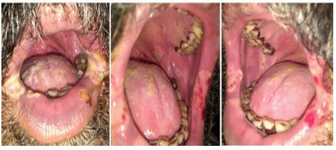
A 60-year male, who hadtested positive for Covid-19,was admitted to our newly started Covid In patient Facility on10 June 2020.The patient was diagnosed based on the Reverse Transcriptase-Polymerised Chain Reaction (RT-PCR) nasopharyngeal swab report. He also haddiabetes and hypertension for which he was taking injectable insulin, and tablet Atorvastatin 10 mg respectively. He was also on Aspirin 75 mg once a day.He was a smoker, an alcoholic, and also had a habit of gutkha (proprietary preparation of areca nut and tobacco) chewing. On 22 June 2020, the patient developed ulcers with yellowishgraypseudo-membrane on either corner of the mouth, associated with bleeding.Several small ulcers were also seen on the buccal mucosa, and the mucosa of the soft and hard palate. These ulcers were ranging from pin point to about 10 mm in dimensions. There was minimal to no erythema immediately surrounding the ulcer nor were there any distinct tissue tags surrounding these ulcerations.White coating was also seen on dorsum of tongue (Figure 1). Ulcerations were thus present on both keratinised and non-keratinised mucosa. Patient had difficulty in chewing, swallowing and talking. A working diagnosis of herpetic ulcers with superimposed candida lesion on bilateral angle of mouth was considered.The patient was prescribed topical anaesthetic and clotrimazole 0.1%. The ulcers started healing by the 3rd day. Subsequently the patient was discharged on 7th July 2020 from the hospital and the ulcers healed completelythereafter as telephonically reported by the patient.
Case 2
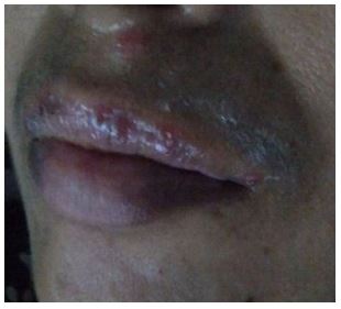
A 50-year asymptomatic female, tested positive for Sars-nCoV-2 on the RT-PCR nasopharyngeal swabwas admitted to the quarantine ward on 13 July 2020. Patient hadno contributory medical history.On 16th July, the patient developed close to ten bilateralvesicles on the upper lip and an erosion close to the nose philtrum (Figure 2). These vesicles subsequently burst and left multiple erosions crossing the midline. There was no evidence of any other lesions in the oral cavity. The patient had difficulty in talking and eating food. A working diagnosis of herpetic vesicular lesion was made.The patient was prescribed topical anaesthetic gel and acyclovir 5% ointment. However, the patient used only the acyclovir gel. The lesions healed completely in the next 4 days. The patient was later discharged on 26th July 2020.

Figure 1: Ulcerative lesions with bleeding areas seen on bilateral corner of mouth covered with slough. Ulcers also seen on the posterior buccal mucosa, hard and soft palate.

Figure 2: Ulcers seen on the upper lip and close to the nose near the philtrum.
Discussion
Oral manifestation reported in COVID-19 are inflammation of wharton’s duct, salivary gland infection, depilation of tongue, xerostomia, candida associated lesions, herpetiform lesions, oral ulcer, apthous ulcers, erythema multiforme type lesions and necrotizing periodontitis [6]. The common site affected are palate, lip, buccal mucosa and tongue [7]. Our first case reported with oral ulcers covered with yellowish pseudomembrane on corner of mouth and small ulcers on buccal mucosa, soft and hard palate also. The oral findings reported in our case series were largely consistent with the findings of the studies reported earlier.Clear relation of COVID-19 and oral mucosal relation has been not established. Some author state that the oral mucosal lesions are due to immunosuppression and stress associated with COVID-19 which causes secondary herpetic infection [6].
Ansari et al [9] had taken biopsy for the oral lesions and the lesions were not attributable to any oral lesion. They had also performed serological tests for HSV 1 and 2 antibodies, which was reported negative. It could therefore be inferred that the ulcers reported by Ansari et al were not herpetic ulcers.
It has been reported that ACE2 is the main host cell receptor of SARS-CoV-2, and plays a crucial role in the entry of virus into the cell,to cause the final infection. Therefore, cells with ACE2 receptor distribution, play hosts for the virus and causes inflammatory reactions in related organs and tissues. Thus, in the oral mucosa too, and specially on the tongue mucosa and salivary glandsare the site for SARS-CoV-2 infection and replication so that such inflammatory reactions may be apparent [7,8]. Surprisingly in our cases, neither the tongue nor the salivary glands seem to be affected.
Etiology and Differential diagnosis
We propose that we should consider the possibility that these lesions could be caused by Sars-nCoV-2 virus directly and not merely represent a secondary manifestation of a drug reaction or an immunosuppressed state.Of course,to validate these theories, there is a need to isolate the Sars-nCoV-2 virus from the oral biopsy specimen of an infected patients using Huh7 and Vero E6 cells [13]. Also isolating the Sars-nCoV-2 virus from the smear obtained from the vesicle could contribute greatly to confirming the role off Sars-nCov 2 virus in its etiology.
One explanation for the oral mucosa getting infected is the sheer proximity of the nasal mucosa and the oral cavity. Another explanation for the same is through frequent touching of the lips by the patient. There were lesions also seen on the buccal mucosa, tongue, soft and hard palate. We postulate that the virus in the droplets from the cough or sneeze of a CoV-2 patient could inoculate the oral mucosa and lead to oral lesions. The above possibilities combined by the expression of ACE2 receptors on oral mucosa and a permeable oral mucosa could be responsible for the oral lesions. It is debatable, given the fact that ACE2 receptors are expressed in the oral mucosa and a permeable oral mucosa, why are oral lesions in most Covid-19 patients? One explanation could be that since the patient is overwhelmed by the general state and its associated stigma, minor oral lesions are ignored by the patient. Only when the oral lesions are alarming will the patient seek attention. This can be seen, in our cases as well. In case1, thepatient sought an Oral Physician’s care only because he was affected with multiple ulcers along with large bilateral lesion on corner of mouth. Case 2 is a parent of our intern and therefore the intern sought expert opinion for her mother even though the symptoms were not severe.
Covid-19 associated dermatological lesions has been reported earlier, with erythematous rash being most common [14]. The lesion on nose philtrum in case 2is the dermatological finding of Covid-19. It is still early to conclude that these ulcers are primarily oral findings of Covid-19 and not, precipitation of viral oral lesions secondary to the immunocompromised state or adverse reaction to several medications taken as part of the treatment for Covid-19. It is also likely that a combination of the three mechanism is responsible for the oral lesions. In future attempt should also be made to correlate the oral findings to the severity of the disease.
Lesion on the corner of mouth of case 1 was ulcerative with superimposed pseudo-membrane as seen in Stevens Johnson syndrome (SJS) and erythema multiforme (EM). The pseudo-membrane coating was considered as candida superimposition. Multiple ulcers in this case resembled herpetic ulcer, though recurrent apthousstomatitis-herpetiformis (RAS-H) was also considered as differential. However unlike RAS-H the lesions were not multiple and also there was no erythematous halo surrounding the lesion.
Our case demonstrated ulcers on both keratinised and non-keratinised mucosa.Reactivation of herpetic lesions because of the immunocompromised state could not be completely ruled out. The finding of pinpoint oral ulcers along with concomitant angular chelitis lesions seem to be apeculiar finding and could beunique to theoral manifestation of COVID-19 virus infection. Interestingly the oral findings on the lip, tongue and other mucosal sites, observed in our case, does not fit into any single differential diagnosis.
The case 2 mainly presented with blisters and erosions mainly on the upper lip. Clinically these ulcers resemble erosions of herpes labialis except that herpes labialis is unilateral. A differential of SJS and EM was also considered in this case. However only upper lip ulcers could suggests contacting these regions with contaminated hands. In both the cases vesicular fluid could not be examined for cytology because of the fear and anxiety that accompanies affliction with this new Corona Virus among patients as well as their primary physicians helping thembattle the disease.
Pathogenesis
A patient infected with Sars-nCoV-2 contains high blood levels of cytokines and chemokines. findings in Some patients have also shown the presence of pro inflammatory cytokines. Cytokines like IL1, IL2, IL8, IL10, TNF-alpha, MCP1, MIP1 alpha has been expressed in other oral mucosal ulcers likerecurrent apthous stomatitis and Bechets syndrome [10,11]. Interestingly in our cases the ulcers on the mobile mucosa clinically did not resembleBechets or apthous ulcers.Additionally, TNF-alpha, IFN-ү, IL2, IL10 is also expressed in Steven Johnson Syndrome (Dodiuk-Gad et al.,2015),one of differential diagnosis in case 1. TNF-alpha has also been attributed to recurrent apthous ulcer in HIV patients [16]. These cytokines are also expressed in Covid-19 patients, thus explaining possible mechanism of ulcer formation in the oral cavity.IL-4, IL-6, IL-8, IFN-ү, and TNF-α has also been expressed at higher levels in oral lichen planus patients [12]. Oral lesions in Patient with Sars-nCoV-2 infections shows histopathologicallyvacuolization exostosis in epithelial layer and, inflammatory cell infiltration, microvascular thrombosis and haemorrhage in mesenchymal layer [7].
However, it is also important to confirm the presence of these cytokines and chemokines from the saliva of the Sars-nCov-2 patients and compare the levels with those of healthy individuals.This is important in case 1 where the ulcers appeared approximately at the time we expect the “cytokine storm” to kick in.However, it is not clear if the same mechanism explains the atypical lesions in the asymptomatic patient in Case 2.
Treatment
Though the general condition of the patient could be quite alarming and little is known regarding the treatment of these patients. Based on our limited experience we believe that the treatment of the oral findingscan be conservative as the lesions are self-limiting. The lesions in our cases healed completely on topical anaesthetic supplemented with topical antifungals and topical antivirals in case 1 and case 2 respectively. It is possible that these lesions are self-limiting. This could be the explanation in case 2. It is also possible that the oral lesions responded to the systemic medications given, as we know that Covid-19 patientsare treated systemically. Therefore, theoral lesionswarrant no major treatment. It is important to note that the information discussed here is based on the limited experience with Covid 19, also the science of both Covid 19 and its associated oral lesions is still evolving.We believe that every new case needs to be reported to identify lesions correctly and in time. We also believe that the following ideas discussed are for all of us to validate or reject by undertaking relevant investigations. Eventually an analysis of many such case reports as well as larger case series on Covid-19 associated oral lesions would help in understanding the oral lesions better.
Conclusion
This case series along with earlier cases suggest that oral lesions could be seen in Covid-19 elderly patients with comorbidities. The lesions present as ulcers on the tongue, lip, buccal mucosa, soft and hard palate. Treatment of these lesions does not seem to be challenging. Dentists needs to be aware of these oral findings and its management.
References
- https://www.worldometers.info/coronavirus/?utm_campaign=homeAdvegas, 2020.
- Zhou P, Yang XL, Wang XG, Hu B, Zhang L, Zhang W, et al. A pneumonia outbreak associated with a new coronavirus of probable bat origin. Nature. 2020; 579: 270-273.
- Li JY, You Z, Wang Q, Zhou ZJ, Qiu Y, Luo R, Ge XY. The epidemic of 2019-novel-coronavirus (2019-nCoV) pneumonia and insights for emerging infectious diseases in the future. Microb. Infect. 2020; 22: 80-85.
- WHO. Clinical Management of Severe Acute Respiratory Infection When Novel Coronavirus (nCoV) Infection Is Suspected. 2020.
- Xu H, Zhong L, Deng J, et al. High expression of ACE2 receptor of 2019-nCoV on the epithelial cells of oral mucosa. Int J Oral Sci. 2020; 12(1): 8. [DOI:10.1038/s41368-020-0074-x].
- Gutierrez-Camacho JR, Avila-Carrasco L, Martinez-Vazquez MC, et al. Oral Lesions Associated with COVID-19 and the Participation of the Buccal Cavity as a Key Player for Establishment of Immunity against SARS-CoV-2. Int J Environ Res Public Health. 2022; 19(18): 11383. [DOI:10.3390/ijerph191811383].
- Silveira FM, Mello ALR, da Silva Fonseca L, Dos Santos Ferreira L, Kirschnick LB, et al. Morphological and tissue-based molecular characterization of oral lesions in patients with COVID-19: A living systematic review. Arch Oral Biol. 2022; 136: 105374. [DOI: 10.1016/j.archoralbio.2022.105374].
- Soares CD, Mosqueda-Taylor A, Hernandez-Guerrero JC, de Carvalho MGF, de Almeida OP. Immunohistochemical expression of angiotensin-converting enzyme 2 in minor salivary glands during SARS-CoV-2 infection. J Med Virol. 2021; 93(4): 1905-1906. [DOI:10.1002/jmv.26723].
- Ansari R, Gheitani M, Heidari F, Heidari F. Oral cavity lesions as a manifestation of the novel virus (COVID-19) [published online ahead of print. 2020. [DOI:10.1111/odi.13465].
- Dalghous AM, Freysdottir J, Fortune F. Expression of cytokines, chemokines, and chemokine receptors in oral ulcers of patients with Behcet's disease (BD) and recurrent aphthous stomatitis is Th1-associated, although Th2-association is also observed in patients with BD. Scand J Rheumatol. 2006;35(6):472-475. doi:10.1080/03009740600905380.
- Tong B, Liu X, Xiao J, Su G. Immunopathogenesis of Behcet's Disease. Front Immunol. 2019;10:665. Published 2019 Mar 29. doi:10.3389/fimmu.2019.00665.
- Humberto JSM, Pavanin JV, Rocha MJAD, Motta ACF. Cytokines, cortisol, and nitric oxide as salivary biomarkers in oral lichen planus: a systematic review. Braz Oral Res. 2018; 32: e82. [DOI:10.1590/1807-3107bor-2018.vol32.0082].
- Helmy YA, Fawzy M, Elaswad A, Sobieh A, Kenney SP, Shehata AA. The COVID-19 Pandemic: A Comprehensive Review of Taxonomy, Genetics, Epidemiology, Diagnosis, Treatment, and Control. J Clin Med. 2020; 9(4): 1225. [DOI:10.3390/jcm9041225].
- Sachdeva M, Gianotti R, Shah M, et al. Cutaneous manifestations of COVID-19: Report of three cases and a review of literature. J Dermatol Sci. 2020; 98(2): 75-81. [DOI:10.1016/j.jdermsci.2020.04.011].
- Dodiuk-Gad RP, Chung WH, Valeyrie-Allanore L, Shear NH. Stevens-Johnson Syndrome and Toxic Epidermal Necrolysis: An Update. Am J ClinDermatol. 2015; 16(6): 475-493. [DOI:10.1007/s40257-015-0158-0].
- MacPhail LA, Greenspan JS. Oral ulceration in HIV infection: investigation and pathogenesis. Oral Dis. 1997; 3(1): S190-S193. [DOI:10.1111/j.1601-0825.1997.tb00358.x].

