Clinical Image - Volume 3 - Issue 1
Parosteal lipoma of the skull
Yannick Canton Kessely*; Aboubacar Aouami; Kader Ndiaye; Brahim Soukaya
Department of Neurosurgery, Renaissance Hospital, Ndjamena, Chad, Senegal.
Received Date : Jan 24, 2023
Accepted Date : Feb 17, 2023
Published Date: Feb 20, 2023
Copyright:© Yannick Canton Kessely 2023
*Corresponding Author : Yannick Canton Kessely, Department of Neurosurgery, Renaissance Hospital, Ndjamena, Chad, Senegal.
Email: kesselycanton@gmail.com
DOI: Doi.org/10.55920/2771-019X/1377
Clinical Image
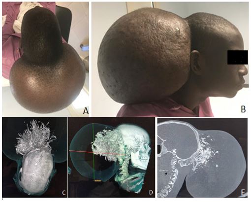
A 17-year-old patient was seen at consultation for a large occipital mass evolving since birth. It was subcentimetric at the beginning and progressively enlarged over the years. Upon consultation, the patient was conscious and had no neurological deficit. The mass was firm, painless and adherent to the deep plane. It was large and measured 21 centimeters anteroposteriorly, 31 centimeters laterally and 25 centimeters craniocaudally (A, B). The cerebral CT scan performed had concluded to a fatty mass with differentiated ossification at the level of its attachment to the external table of the diploea not causing bone lysis. There was no evidence of malignancy. No endocranial anomaly was reported. There was fibrous dysplasia of the sphenoid and bony remodeling of the right lamina and articular process of the 2nd and 3rd cervical vertebrae (C, D, E). Exeresis of the mass was performed. The diagnosis of parosteal lipoma was made. Complete surgery remains the best treatment. Parosteal lipoma of the skull is a very rare benign tumor.


