Clinical Image - Volume 3 - Issue 1
kerion in an elderly patient
Hashas FZ*; Douhi Z; Chhiti S; Soughi M; Elloudi S; Bay Bay H; Mernissi FZ
Department of Dermatology and Venereology, University Hospital Hassan II, Route Sidi Hrazem, Faculty of Medicine and Pharmacy, Sidi Mohamed Ben Abdellah University, Fez, Morocco.
Received Date : Jan 28, 2023
Accepted Date : Feb 21, 2023
Published Date: Feb 23, 2023
Copyright:© Hashas fatima zahra 2023
*Corresponding Author : Hashas fatima zahra, Department of Dermatology and Venereology, University Hospital Hassan II, Route Sidi Hrazem, Faculty of Medicine and Pharmacy, Sidi Mohamed Ben Abdellah University, Fez, Morocco.
Email: hashas.fz@gmail.com
DOI: Doi.org/10.55920/2771-019X/1380
Clinical Image
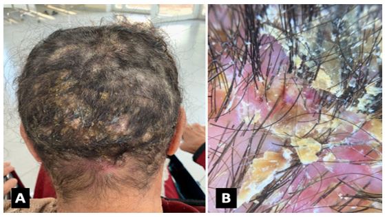
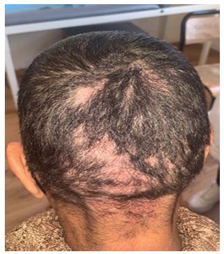
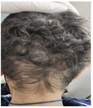
Tinea capitis is generally considered a common condition in children, but not in adults. If the infection occurs in adults, it may have an atypical appearance. We present an elderly woman with inflammatory tinea capitis. A 71-year-old rural woman presented with a 2-month history of multiple pruritic, crusted, and pustular lesions on the scalp. She had been treated with antibiotics without any improvement. The patient was not on any immunosuppressant drug and no other family member was suffering from similar disease however an animal contacting history was found . The patient had no fever or other constitutional symptoms. Physical examination revealed an elderly woman in otherwise good general condition. The scalp showed multiple, crusted plaques involving the occipital area of scalp. The lesions were non- indurated, mildy tender and studded with numerous pustules. A non-scary alopecia was observed in and around the lesions. The hairs present were easily plucked and matted. Dermoscopic findings were : greasy scales, perifollicular white macules and bent hairs (Figure 1). The posterior cervical lymphnodes were enlarged and palpable. A potassium hydroxide (KOH) preparation made from scalp and hair scrapings showed fungal hyphae. The diagnosis of kerion was made and the patient was treated with oral and topical griseofulvin for eight weeks. There was complete clearance of lesions hair regrowth (Figures 2,3).

Figure 1: Clinical and dermoscopic features of Tinea capitis A- Erythematous pseudoalopecic plaques with yellow crusts involving the occipital area of scalp. B- Greasy scales, bent hairs.

Figure 2: Evolution after one month of treatment.

Figure 3: Evolution after three months of treatment.
References
- Gianni C, Betti R, Perotta E, Crosti C. Tinea capitis inadults. Mycosis. 1995; 38:3 29-31.
- Anstey A, Lucke TW, Philpot C. Tinea capitis caused by Trichophyton rubrum. Br J Dermatol. 1996; 135: 113-5.
- Ziemer A, Kohl K, Schroder G. Trichophyton rubrum induced inflammatory tinea capitis in a 63-year-old man. Mycosis. 2005; 48: 76-9.

