Case Report - Volume 3 - Issue 2
Concomitant Bullous and Crusted Scabies: A Rare Presentation
Sektaoui S*; Mehsas Z; Meziane M; Senouci K
Department of dermatology & venereology, Ibn Sina University Hospital, Faculty of medicine and pharmacy of rabat, Mohamed V University, Rabat, Morocco.
Received Date : Feb 03, 2023
Accepted Date : Mar 13, 2023
Published Date: Mar 20, 2023
Copyright:© Sektaoui S 2023
*Corresponding Author : Sektaoui S, Department of dermatology & venereology, Ibn Sina University Hospital, Faculty of medicine and pharmacy of rabat, Mohamed V University, Rabat, Morocco.
Email: soukaina.sektaoui@gmail.com
DOI: Doi.org/10.55920/2771-019X/1398
Abstract
Scabies is a highly contagious skin condition caused by an infestation of the mite Sarcoptes scabiei. The mites burrow into the upper layers of the skin and lay their eggs, causing intense itching and characteristic linear burrows. There are several atypical clinical variants of scabies, including bullous, crusted, hidden, incognito, nodular, and scalp scabies. Bullous scabies is an uncommon variant, characterized by the formation of large, fluid-filled blisters (bullae) on the skin. Crusted scabies, also known as Norwegian scabies, is a severe and highly contagious form of scabies characterized by thick, crusted lesions on the skin. In this case report, we present a rare case of a 69-year-old man with concomitant bullous and crusted scabies. The patient was treated with a whole-body application of benzyl benzoate lotion and two doses (12mg) of oral ivermectin, two weeks apart, and the skin lesions disappeared 20 days later. This case highlights the importance of considering scabies as a differential diagnosis in patients presenting with bullous lesions and the need for careful examination and investigation to distinguish between bullous scabies and bullous pemphigoid.
Keywords: Scabies, Bullous Scabies, Crusted Scabies, Bullous Pemphigoid, Mite Infestation, Skin Infestation.
Introduction
Human scabies is a highly contagious skin condition caused by an infestation of the mite Sarcoptes scabiei. The mites burrow into the upper layers of the skin and lay their eggs, causing intense itching (pruritus) and characteristic linear burrows. The most common symptoms of scabies include intense itching, especially at night, and a rash with small red bumps and blisters. There are several atypical clinical variants of scabies, including bullous, crusted, hidden, incognito, nodular, and scalp scabies. Bullous scabies is an uncommon variant, characterized by the formation of large, fluid-filled blisters (bullae) on the skin. These blisters can closely resemble the autoimmune blistering disorder bullous pemphigoid in appearance. Crusted scabies, also known as Norwegian scabies, is a severe and highly contagious form of scabies characterized by thick, crusted lesions on the skin. It is most commonly seen in individuals with weakened immune systems, such as those with HIV/AIDS or other chronic illnesses. In this case report, we present a rare case of a 69-year-old man with concomitant bullous and crusted scabies.
Case report
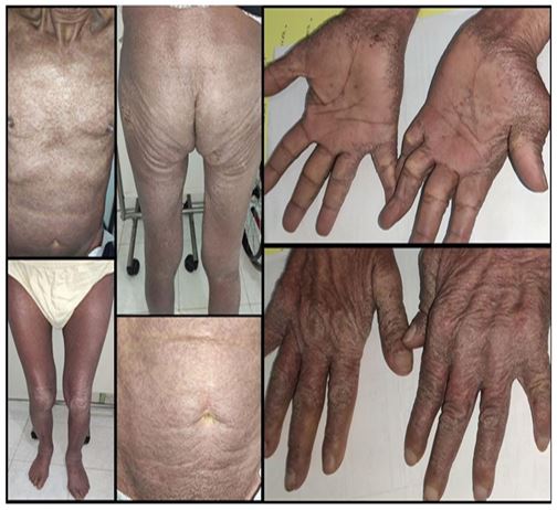
A 69-year-old man, who had a history of benign prostatic hyperplasia and cervical trauma, was referred to our department for a 6-month history of generalized itchy, papular and scaly eruptions that spared the face. Despite treatment with oral antihistamines and corticosteroids, as well as a combined topical corticosteroid and petrolatum emollient, the itching persisted. Laboratory tests revealed significant eosinophilia, and a physical examination revealed exfoliative erythroderma with multiple scratching lines on the back and trunk, as well as scabies burrows on the hands and wrists (figure 1). A diagnosis of scabies was confirmed through the microscopic examination of skin scrapings. The patient was treated with a whole-body application of benzyl benzoate lotion and two doses (12mg) of oral ivermectin, two weeks apart.
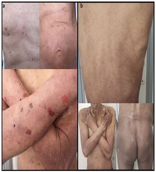
However, two weeks later, the patient developed multiple tense blisters on the trunk and abdomen, and post-bullous erosion on the arms (figure 2a). A biopsy and histopathology of the blisters revealed a subepidermal blister filled with serous fluid and an inflammatory infiltrate composed of lymphocytes, neutrophils, and eosinophils. We also found eosinophils in the papillary dermis at the base of the blister. Both direct immunofluorescence microscopy and the value of anti-basement membrane zone (BMZ) antibodies were negative. The diagnosis of bullous scabies was made, and the patient received two additional doses of oral ivermectin, two weeks apart. Twenty days later, the patient's skin lesions had completely disappeared as shown in figure 2b.

Figure 1: This clinical image illustrates the presence of exfoliative erythroderma on the patient's back and trunk, characterized by multiple scratching lines. Additionally, scabies burrows can be seen on the patient's hand, between the fingers, in the flexure of the wrist, and on the palm, providing visual evidence of a scabies infestation.

Figure 2: This clinical image (a) depicts the patient’s condition two weeks after the initial treatment, displaying multiple tense blisters on the trunk and abdomen, as well as post-bullous erosion on the arms. Image (b) shows the final outcome of the treatment, where the skin lesions have completely disappeared. This illustrates the progression of the bullous scabies and the effectiveness of the chosen treatment regimen.
Discussion
Scabies, a cutaneous parasitosis caused by the mite Sarcoptes scabiei hominis, exists in two forms: common and crusted scabies. Common scabies is benign and moderately contagious, while crusted or Norwegian scabies is more severe, highly contagious, and often associated with immunosuppression [1]. Bullous scabies is a rare presentation of the mite infestation, with only 46 reported cases in the literature. However, only one case of concomitant bullous and crusted scabies has been described by Wen-Jing Su et al. in 2015 [2]. Diagnosis of scabies can be challenging, particularly when the eruption is treated with glucocorticoid therapy, as this can exacerbate the scabies and cause blisters to develop from the original skin lesions [3]. The presence of blisters in scabies is exceptional and multiple mechanisms have been proposed, including superinfection of mite lesions with Staphylococcus aureus [3], damage to the anti-basement membrane zone (BMZ) by the mite's lytic enzymes [4], the production of autoantibodies in response to cross-reacting mite components with bullous pemphigoid antigen [5], autoeczematisation to the scabies mite or an immunity response type 1 to an antigen from the saliva of the scabies [6]. A wide variety of symptoms can occur in scabies, and the presence of bullae is extremely rare. However, patients with scabies and bullae are often misdiagnosed as having bullous pemphigoid. This can be difficult to distinguish, especially when skin scrapings do not show any mites or eggs. However, there are some differentiating features between these two entities that can be used to make a diagnosis: Bullous scabies can affect individuals of all ages, while bullous pemphigoid primarily affects older individuals. Additionally, bullous pemphigoid always shows linear C3 or IgG deposition in the basement membrane zone (BMZ), while bullous scabies may show both linear and granular deposition in the BMZ [7]. Scabies is contagious and a family history is very important. In cases of bullous pemphigoid, good responses are seen after oral prednisolone administration, while bullous scabies may persist and need to be treated with antiscabies drugs like Ivermectin [8, 9].
Conclusion
In conclusion, this case report highlights the importance of considering scabies as a differential diagnosis in patients presenting with tense bullous lesions associated with pruritus and maculopapular rash, specifically if good responses are not seen after steroid treatment. To prevent misdiagnosis as bullous pemphigoid, scrapings for Sarcoptes scabiei mites and eggs should be done as soon as possible.
Acknowledgements
Sektaoui Soukaina and Mehsas Zoubida participated in the research design, the writing of the paper. Pr Meziane Meriem and Senouci Karima participated in the research design
Funding: The authors declare no funding
Disclosure: The authors declare no conflicts of interest
Author contributions:
- Dr Sektaoui Soukaina : participated in the research design, the writing of the paper
- Dr Mehsas Zoubida: participated in the research design, the writing of the paper
- Pr Meziane Meriem: participated in the research design
- Pr Senouci Karima: participated in the research design
References
- Owersey L, Cunha MX, Feldman CA et al. Dermoscopy of Norwegian scabies in a patient with acquired immunodeficiency syndrome. An Bras Dermatol. 2010;85(2):221-223.
- Su WJ, Fang S, Chen A et al. A case of crusted scabies combined with bullous scabies. Exp Ther Med. 2015;10(4):1533-1535.
- Ansarin H, Jalali MH, Mazloomi S et al. Scabies presenting with bullous pemphigoid-like lesions. Dermatol Online J. 2006;12(1):19.
- Veraldi S, Scarabelli G, Zerboni R et al. Bullous scabies. Acta Derm Venereol. 1996;76(2):167-168.
- Ostlere LS, Harris D, Rustin MH. Scabies associated with a bullous pemphigoid-like eruption. Br J Dermatol. 1993;128(2):217-219.
- Maan MA, Maan MS, Sohail AM et al. Bullous scabies: a case report and review of the literature. BMC Res Notes. 2015;8:254.
- Salo OP, Reunala T, Kalimo K et al. Immunoglobulin and complement deposits in the skin and circulating immune complexes in scabies. Acta Derm Venereol. 1982;62(1):73-76.
- “Scabies prevention and control guidelines acute and subacute care facilities” by los angeles county department of public health acute communicable disease program
- Mir F, Cruz-Oliver DM. Scabies manifesting as bullous pemphigus in a nursing home resident. J Am Geriatr Soc. 2014;62(6):1201-1203.

