Clinical Image - Volume 3 - Issue 2
Atoll sign : l’éventail diagnostic
Mribat M*; El Adioui G; Rouni L; Laamrani Fz
Central Radiology Department, CHU Ibn Sina, Mohamed V University, Rabat, Morocco
Received Date : Feb 17, 2023
Accepted Date : Mar 16, 2023
Published Date: Mar 23, 2023
Copyright:© Mribat M 2023
*Corresponding Author : Mribat M ,Central Radiology Department, CHU Ibn Sina, Mohamed V University, Rabat, Morocco
Email: meriememribat@gmail.com
DOI: Doi.org/10.55920/2771-019X/1401
Clinical Image
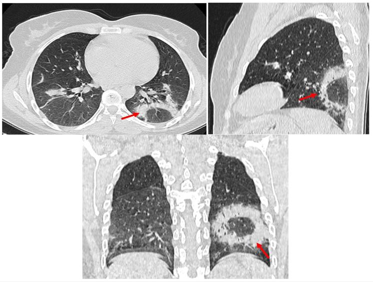
The atoll sign or inverted halot sign is seen on high-resolution chest CT scans as a central frosted glass area surrounded by peripheral condensation in a ring or crescent, the contours of which may be smooth or spiculated [1]. Histologically, the central zone (ground glass opacity) corresponds to alveolar septal inflammation and cellular debris in the alveolar spaces, while the condensed peripheral zone corresponds to granulomatous tissue in the distal air spaces [2]. Long considered pathognomonic for cryptogenic organizing pneumonia (COP). Currently, the atoll sign is found in other pathologies such as vasculitis (Wegener's disease), sarcoidosis, pneumocystosis, paracoccidioidomycosis, tuberculosis, lipid pneumopathy or after radiofrequency treatment… [3] Recently, this sign was described among the atypical appearances on chest CT scan in patients with covid 19 infection. [4]

Figure 1: Chest CT (3 planes) showing a typical image of an inverted halo “atoll sign” (arrow).
AML should be differentiated from other cystic lesions of the lung, mainly histiocytosis X; which affects the smoking subject with a characteristic appearance on the scanner of a cyst of irregular lace shapes, predominant in the middle and upper lobe, but the distinction sometimes remains difficult in the advanced stage of the disease, and also with lymphocytic interstitial pneumonia (LIP) which is characterized by ground glass patches around the cysts, distributes around the bronchovascular sheaths. Although the chest CT scan may be compatible or characteristic of AML, the definitive diagnosis is based on the combination of clinico-radiological criteria and the histological confirmation obtained by lung biopsy or biopsy of a lymph node or lymphangiomyoma [6].
References
- Hansell DM,et all.Fleischner Society: glossary of terms for thoracic imaging.Radiology 2008;246 (3):697-722.
- Voloudaki AE, Bouros DE, Froudarakis ME et-al. Crescentic and ring-shaped opacities. CT features in two cases of bronchiolitis obliterans organizing pneumonia (BOOP). Acta Radiol. 1997;37 (6): 889-92.
- Marchiori E et all.Reversed halo sign : high-resolution CT scan findings in 79 patients. Chest 2012;141 (5):1260-6.
- B Lodé et all. Imaging of covid 19 pneumonia. JIDI 2020

