Clinical Image - Volume 3 - Issue 2
Uterine artery embolization for the management of vaginal bleeding in endometrial cancer and ACS condition
Azin A MD, FASE, FACC1; Kiara R2; Mina M1; Alia B1; Masoud S1; Kamran R3; Azam Y MD1*; Koroosh K4
1Cardio-Oncology Research Center, Rajaie Cardiovascular Medical and Research Center, Iran University of Medical Sciences, Tehran, Iran.
2Rajaie Cardiovascular Medical and Research Center, Iran University of Medical Sciences, Tehran, Iran.
3Cancer Institute, Imam Khomeini Hospital, Tehran University of medical Sciences, Tehran, Iran
4Interventional cardiology Research Center, Rajaie Cardiovascular Medical and Research Center, Iran University of Medical Sciences, Tehran, Iran.
Received Date : Feb 17, 2023
Accepted Date : Mar 20, 2023
Published Date: Mar 27, 2023
Copyright:© Azam Y 2023
*Corresponding Author : Azam Y, Cardio-Oncology Research Center, Rajaie Cardiovascular Medical and Research Center, Iran University of Medical Sciences, Tehran, Iran.
Email: Cardiooncology.r@gmail.com
DOI: Doi.org/10.55920/2771-019X/1403
Clinical Image
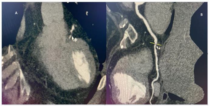
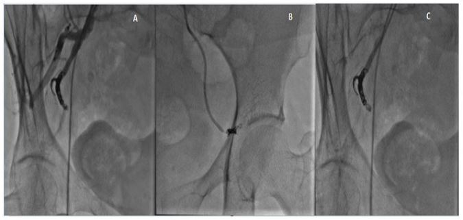
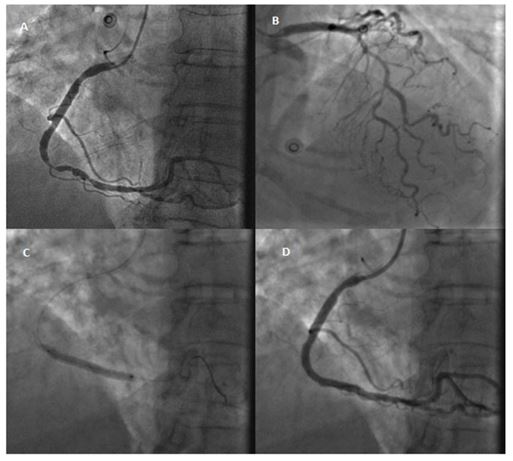
A 76-year-old female with a history of hypothyroidism, arterial hypertension, Dyslipidemia and diabetes mellitus type 2 presented with vaginal bleeding that is diagnosed endometrial cancer for her. She had exertional chest pain function class 3. Coronary CT angiography was done and multiple significant lesions in mid part of the right coronary artery( RCA) and mid part of the left anterior descending artery( LAD) were noted. Exertional chest pain with optimal medical treatment does not resolve and due to vaginal bleeding and contraindication of antiplatelet therapy, the patient underwent angioemboliFigure 1: Coronary CT Angiography shows A .significant lesion mid part LAD , B. multiple significant lesions mid part RCA. zation of both right iliac and uterine arteries with micro coils and PVA 350-500 microns . The procedure was done successfully and vaginal bleeding stopped. After three days, the patient underwent coronary angiography via right radial artery and PCI on RCA was done with ultimaster 3.5*38 mm DES with a good final result. Dual antiplatelet therapy (DAPT) was prescribed for her and no bleeding occurred in the 3 months’ follow-up. Also, the patient did not have any episodes of chest pain or cardiovascular problems. Chemotherapy was started and continued for the patient without significant complications.

Figure 1: Coronary CT Angiography shows A .significant lesion mid part LAD , B. multiple significant lesions mid part RCA.

Figure 2: Angio embolization of A,B.right iliac artery, C. right uterine artery with micro coils and PVA 350-500 microns.

Figure 3: Coronary angiography shows A. multiple significant lesions midpart of RCA, B.significant lesion mid part LAD , C. PCI on RCA with ultimaster 3.5*38 mm DES, D.RCA after angioplasty with good result.

