Clinical image - Volume 3 - Issue 2
Myelinated Retinal Nerve Fibers
Taouri N*; Baiz T; H’meimett Z; Benchekroun S; Boutmzine N; Amazouzi A; Cherkaoui Lo
Mohammed V University of Rabat Faculty of Medicine and Pharmacy of Rabat, Morocco.
Received Date : Mar 06, 2023
Accepted Date : Mar 31, 2023
Published Date: April 07, 2023
Copyright:© Taouri N 2023
*Corresponding Author : Taouri N, Mohammed V University of Rabat
Faculty of Medicine and Pharmacy of Rabat, Morocco.
Email: ophtalmo-taouri@outlook.fr
DOI: Doi.org/10.55920/2771-019X/1412
Clinical image
We report a case of a 25-year-old-woman, with no pathological history. The patient presented to ophthalmology consultation for a routine eye examination.
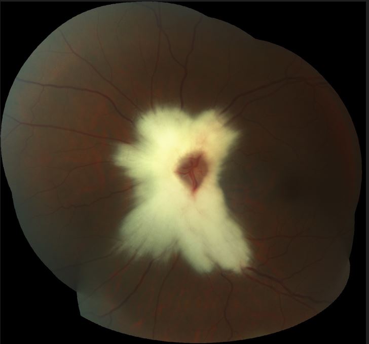
Her Uncorrected visual acuity was 10/10 for both eyes. On slit lamp examination, anterior segment was normal, while fundus examination was found to have on the left eye, a flat grayishwhite area with irregular borders concentric to the optic disc. Otherwise the posterior segment examination of left eye was normal. Our diagnosis was myelinated retinal nerve fibers. the first clinical description of myelinated retinal nerve fibers was in1856 by Virchow[1]. According to several authors: it corresponds to myelination of retinal nerve fibers, which are normally devoid of myelin. It is represented clinically as white to grey patch following the distribution of the nerve fibres , with irregular feathery borders. The area of myelinesation can be concentric to the optic disc or in periphery retina[1, 2].
several studies report that is due to dysfunction of the lamina cribrosa and to ectopic oligodendrocytes[2].
Also, previous studies concerning clinical and pathologic features have reported that it is a congenital anomaly that occur in 0,5% to 1% of population[1,3].usually asymptomatic and unilateral, and can be associated with myopia and amblyopia. [2, 4,5,6]
Conflict of interest: The author declares that there is no conflict of interest.
Figure 1: Fundus photography showing a thick bunch of myelin fibers concentric to the optic disc.

References
- Grzybowski A, Winiarczyk I: Myelinated retinal nerve fibers (MRNF) - dilemmas related to their influence on visual function. Saudi J Ophthalmol. 2015, 29:85-8. 1
- Tarabishy AB, Alexandrou TJ, Traboulsi EI: Syndrome of myelinated retinal nerve fibers, myopia, and amblyopia: a review. Surv Ophthalmol. 2007, 52:588-96
- Straatsma BR, Foos RY, Heckenlively JR, Taylor GN. Myelinated retinal nerve fibers. Am J Ophthalmol. 1981; 91(1):25-38.
- Lam AK, Pang PC. The effect of myelination on perimetry and retinal nerve fibre analysis. Clin Exp Optom. 2000; 83(1):4-11.
- Holmes JM, Clarke MP: Amblyopia. Lancet. 2006, 367:1343-51. 10.1016/S0140-6736(06)68581-4
- Holmes JM, Beck RW, Kraker RT, et al.: Impact of patching and atropine treatment on the child and family in the amblyopia treatment study. Arch Ophthalmol. 2003, 121:1625-32

