Clinical Image - Volume 3 - Issue 2
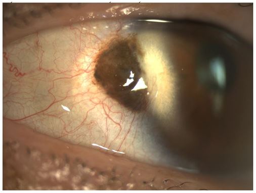
Conjunctival nevus superimposed on a Pterygium, with a possible corneal involvement: A challenging case
Saoiabi Y*; Bouirig K; Cherkaoui O
Mohammed V University of Rabat, Specialty Hospital of Rabat, Morocco.
Received Date : Mar 19, 2023
Accepted Date : April 13, 2023
Published Date: April 20, 2023
Copyright:© Yahya Saoiabi 2023
*Corresponding Author : Yahya Saoiabi, Mohammed V University of Rabat, Specialty Hospital of Rabat, CHU Ibn Sina, Morocco.
Email: Saoiabi.yahya@gmail.com
DOI: Doi.org/10.55920/2771-019X/1421
Clinical Image
The presented photograph shows a suspicious lesion of conjunctival nevus in a 42-year-old patient. The patient reported that the lesion has been present since childhood and she does not believe that it has increased in size. The lesion is located near the temporal limbus and is superimposed on an early pterygium. There is also evidence of corneal invasion, possibly by the pterygium or the lesion. The lesion measures approximately 5 mm x 6 mm in diameter, is brownish in color, and presents irregular pigmentation and slight elevation. Although the lesion is asymptomatic, it was considered suspicious due to its atypical features and the potential for corneal invasion. Given the proximity to the limbus and the potential for corneal involvement, excision of the lesion and pterygium should be considered. Close monitoring of the patient is also recommended to ensure that there is no recurrence or further corneal involvement. High-resolution imaging of the lesion, such as anterior segment optical coherence tomography (OCT) or ultrasound biomicroscopy (UBM), can be useful to better characterize its morphology and assess for any deeper invasion. The management plan should be individualized for each patient based on their specific risk factors and comorbidities. Management of conjunctival nevi is determined by several factors such as size, location, and the risk of malignancy. Small and asymptomatic nevi can be observed with regular followup. However, large and symptomatic nevi may require surgical excision. The surgical approach will vary depending on the location and extent of the lesion.
Histological examination of the excised tissue is critical in determining if the nevus shows features indicative of malignancy or high risk for malignancy. If malignancy is present, additional excision, sentinel lymph node biopsy, or systemic evaluation for metastatic disease may be necessary.


