Clinical Image - Volume 3 - Issue 3
Thimble Bladder
Sanjeev Sharma1; Rajesh Chaudry2; Manjeet Kumar3*
1Postgraduate MS Scholar (AMO), Department of Shalya Tantra, Rajiv Gandhi Government Post-Graduate Ayurvedic College Paprola, Kangra (HP), India.
2Assistant Professor, Department of Surgery, Pandit Jawahar Lal Nehru Government Medical College Chamba, HP, India.
3Assistant Professor, Department of Urology, IGMC, Shimla HP, India
Received Date : Mar 26, 2023
Accepted Date : April 27, 2023
Published Date: May 04, 2023
Copyright:© Manjeet Kumar 2023
*Corresponding Author : Manjeet Kumar, Assistant Professor, IGMC Shimla HP, India.
Email: dr.vicky.surgeon@gmail.com
DOI: Doi.org/10.55920/2771-019X/1431
Clinical Image
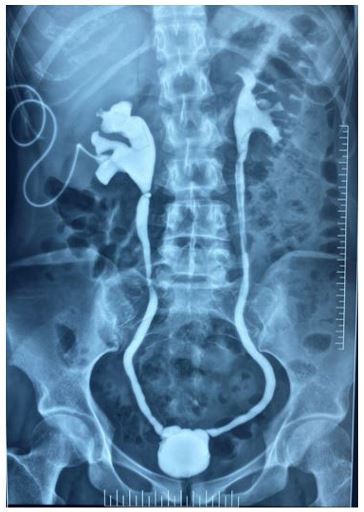
Thimble bladder is a small, contracted, thick urinary bladder after chronic infection, inflammation, and fibrosis following genitourinary tuberculosis. The bladder is involved when tubercle bacilli are excreted in urine from the Kidneys. Small capacity urinary bladder presents with frequency, incontinence, and deranged creatinine.
A 28-year female presented with frequency of micturition, dysuria, and low-grade fever. The urine culture was sterile with 18-20 pus cells/ HPF. Urine for AFB was positive. Ultrasound abdomen showed bilateral hydronephrosis with decreased urinary bladder capacity. A bilateral percutaneous nephrostomy was done. Cystogram was suggestive of tortuous ureters, small capacity thimble urinary bladder.
She was started on antitubercular therapy with Isoniazid, Rifampicin, ethambutol, and pyrazinamide for 6 months. Urine for AFB was negative at 3 months. She underwent simple cystectomy and ileal conduit as urinary bladder was small (25 ml) in capacity. Hans Wildbolz coined term Genitourinary tuberculosis (GUTB) in 1937. Dissemination of tubercle bacilli from pulmonary focus leads to GUTB.
Genitourinary tuberculosis is second most common extrapulmonary tuberculosis in developing countries, whereas it is third most common in developed countries. It accounts for 20-40% of all cases of extra pulmonary tuberculosis (EPTB) cases [1]. GUTB most commonly involves kidney (74% cases) followed by epididymis, testis, bladder, ureter and prostate gland. Isolated genital organ involvement is reported in 5-30% of tuberculosis cases [2,3]. Non-functioning kidneys, ureteric stricture and small capacity urinary bladder are delayed consequences of untreated GUTB [4]. Treatment of small capacity urinary bladder is augmentation cystoplasty; in case of thimble bladder simple cystectomy and diversion is treatment of choice.

Case study 3
A 76-year-old male with a past medical history of peripheral arterial disease (PAD) presented with a necrotic ulcer to the lateral aspect of the 5th digit following a previous gangrenous ulcer and secondary to metatarsal head resection of the 5th digit. Due to the prior PAD diagnosis, the patient required revascularization but refused vascular treatment. Limited vascular perfusion to the ulcerative site relieved the chronic nature of the ulcer. Prior to the first application of ActiGraft, the wound appeared chronic with gangrenous and ischemic changes. The ulcerative site measured approximately 15 cm2 with a fibronecrotic wound bed (Figure 3A). Cellulitic changes were noted to the foot with noted undermining and tunneling. The patient underwent two applications of the autologous blood clot applied a week apart (Figure 3B). Following the first application, there was an improvement in the ulcerative site, and the wound bed was reduced to approximately 5 x 2 cm (Figure 3C). Furthermore, there was a decrease in the fibronecrotic wound bed with increased granulation to the ulcerative site and re-epithelialization of the peri-wound area/edges. Following the second application, there was sustained healing of the ulcerative site with a reduction in size to approximately 4 x 1.7 cm, reduced undermining, and continued re-epithelialization of the surrounding wound edges (Figure 3D). Follow-up visits showed increased re-epithelialization of the peri-wound area with continuous granulation of the wound site. The ulcerative site measured approximately 2.9 x 1.1 cm (Figure 3E).
Case study 4
A 40-year-old female with a past medical history of meningocele (previous procedure for correction) and Charcot neuroarthropathy presented to the clinic following the development of an ulcer to the lateral aspect of the right foot. Offloading via a Charcot Restraint Orthotic Walker (CROW) boot was used, and the patient underwent a reconstructive pedal surgery to treat osseous and joint destruction and prevent further breakdown of the soft tissue and the spread of infection. Although various wound care modalities (i.e., multiple debridements, Medical-honey, collagen, hyaluronic acid, Negative Pressure Wound Therapy (NPWT), and PRF (platelet-rich fibrin)) were tried, granulation and re-epithelialization of the wound bed were not achieved. Prior to the first application of ActiGraft, the ulcer measured approximately 2.8 cm2 and exhibited a fibrogranular wound bed (Figure 4A).
References
- Kulchavenya E. Best practice in the diagnosis and management of urogenital tuberculosis. Ther Adv Urol. 2013; 5:143-151.
- Yadav S, Singh P, Hemal A, Kumar R. Genital tuberculosis: current status of diagnosis and management.Transl Androl Urol. 2017; 6(2): 222-33.
- Manso MC, Rodeia SC, Rodrigues S, Domingos R. Synchronous presentation of two rare forms of extrapulmonary tuberculosis. BMJ Case Rep. 2016; 18: 10.
- Raina P, Sharma GK, Kumar M, Barwal KC, Rana K, Chauhan S. Genitourinary tuberculosis: clinical profile, diagnostic approach and treatment outcome in a tertiary care center of North India. International Journal of Health and Clinical Research. 2021; 4(8): 138-142.

