Case report - Volume 3 - Issue 3
Retroperitoneal Bronchogenic Cyst with Intracystic Hemorrhage Masquerading as an Adrenal Tumor: A Rare Presentation
Anshuman Singh1, Kasi Viswanath Gali1, Vivek Pai1, Bhavna Nayal2, Padmaraj Hegde1*
1Department of Urology, Kasturba Medical College, Manipal, Manipal Academy of Higher Education (MAHE), Manipal (KA), India.
2Department of Pathology, Kasturba Medical College, Manipal, Manipal Academy of Higher Education (MAHE), Manipal (KA), India.
Received Date : Mar 31, 2023
Accepted Date : May 02, 2023
Published Date: May 09, 2023
Copyright:© Padmaraj Hegde 2023
*Corresponding Author : Padmaraj Hegde, Professor, Department of Urology, Kasturba Medical College, Manipal, Manipal Academy of Higher Education, Manipal (KA), India
Email: padmaraj.hegde@manipal.edu
DOI: Doi.org/10.55920/2771-019X/1434
Abstract
Bronchogenic cysts are primitive, foregut-derived developmental anomalies with bronchial-type, pseudostratified cylindrical epithelium commonly occurring in the thorax. Bronchogenic cysts can occur in the retroperitoneum, albeit rarely. We report a case of a 53-year-old man presenting with vague abdominal pain who was diagnosed with a well-defined lesion in the left supra renal region on computed tomography imaging with few haemorrhagic, calcific, and heterogeneously enhancing foci. The left adrenal gland could not be delineated separately. Preoperative workup for a functioning adrenal tumour was negative. The differential diagnoses were atypical adrenal cyst, neurogenic ganglioma, and exophytic diaphragmatic tumour. The patient underwent excision of the left adrenal/retroperitoneal mass. Histopathology revealed features suggestive of a bronchogenic cyst. Diagnostic imitations exist preoperatively in a bronchogenic cyst, and only histology can provide a definitive diagnosis. Surgery remains the treatment of choice.
Introduction
Bronchogenic cysts are congenital abnormalities of the tracheobronchial bud originating from the primitive foregut, commonly found in the thorax. Occurrence of such cysts in the retroperitoneum is rare [1], with the common site of occurrence being the corpus of pancreas or left adrenal gland [2].
Bronchogenic cysts are commonly asymptomatic, detected incidentally on imaging unless they are infected or enlarged enough to compress nearby organs. Although imaging modalities can help detect these cysts, only histopathological examination can provide a definitive diagnosis of retroperitoneal bronchogenic cyst. Hence, surgical resection remains the treatment of choice to establish definitive histology and provide symptom resolution.
Case report
A 53-year-old male presented with poorly localized abdominal pain for over six months. Systemic examination was normal. Contrast-enhanced CT scan of the abdomen revealed a well-defined lesion in the left supra renal region measuring 7.7 x 6.1 x 9.5cm (AP x TR x CC) with few haemorrhagic, calcific, and heterogeneously enhancing foci within (figures 1-3). The left adrenal gland could not be delineated separately
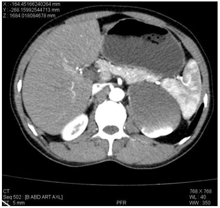
Figure 1: CECT abdomen and pelvis – Axial film; showing left suprarenal cystic lesion with calcific foci and areas of heterogenous enhancement.
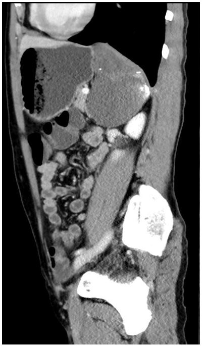
Figure 2: CECT abdomen and pelvis – Sagittal film; showing left suprarenal cystic lesion with calcific foci and areas of heterogenous enhancement.
The lesion was seen abutting the left hemidiaphragm superomedially and extended up to the hiatus with infiltration into the left crus. aboratory parameters were normal. The secretory levels of all adrenal hormones were within normal limits. The working differential diagnoses were atypical adrenal cyst, neurogenic ganglioma and exophytic diaphragmatic tumour. On surgical exploration, a cyst appearing to arise from the left adrenal gland, densely adherent to the inferior surface of the left diaphragm, was noted. Left adrenalectomy with cyst excision in toto was done. Postoperative period was uneventful, and the patient was discharged on the fourth postoperative day. On microscopic examination of the specimen, multiple cysts lined by ciliated pseudostratified columnar epithelium, smooth muscle bundles, seromucinous glands, hyaline cartilage and focal bony formation were identified, leading to the histopathological diagnosis of a bronchogenic cyst (Figure 4-5). Microscopy also revealed abundant hemosiderin-laden macrophages, lymphoid aggregates, and congested vessels suggestive of haemorrhage/infection within the cyst.
Discussion
The tracheobronchial tree's abnormal budding results in benign congenital foregut malformations known as bronchial cysts. Bronchogenic cysts are primarily unilocular or oligo-locular in appearance under the microscope. They are lined with pseudostratified ciliated columnar epithelium that contains bronchial glands, cartilage, smooth muscle, and mucoid material (3). Most frequently, bronchogenic cysts are located in the mediastinum of the thorax (4).
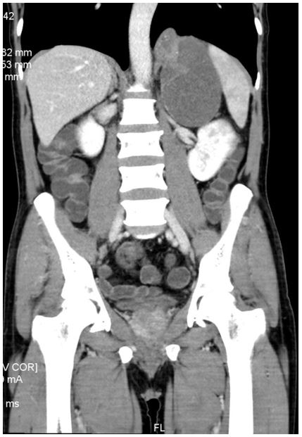
Figure 3: CECT abdomen and pelvis – Coronal film; showing left suprarenal cystic lesion with calcific foci and areas of heterogenous enhancement.
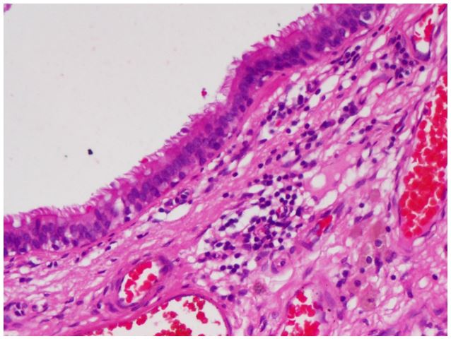
Figure 4: Cyst wall lined by respiratory epithelium (H&E, x200).
Since only a few of these cysts have been found in the retroperitoneal space, they are thought to be rare. Miller et al. first described retroperitoneal bronchogenic cysts in 1953 [5]. Most of these cysts have a diameter of less than 5 cm and are located in the left adrenal gland or pancreatic body [6]. Cysts are typically discovered incidentally and without any symptoms. However, if clinical symptoms are present, they typically result from
secondary bleeding, infection, perforation, or compression of nearby organs and include nausea, vomiting, and poorly localized abdominal pain [7]. This may help to explain the symptoms of abdominal pain that we experienced in our case, which was likely brought on by an infection or haemorrhage inside the cyst.
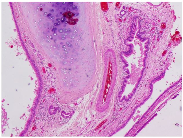
Figure 5: Cyst wall lined by respiratory epithelium overlying stroma showing cartilage and congested vessels. (H&E, x40).
CT is frequently used to detect bronchogenic cysts, which appear as heterogeneous or homogeneous hypoattenuating lesions that are not enhanced by intravenous contrast [9]. In conditions of haemorrhage, however, thick mucinous secretions of the lesions frequently appear as enhancing areas within the cyst [10]. In addition, calcification is more prevalent in bronchial cysts, and it is believed to be the primary contributor to high attenuation on unenhanced CT scans [11]. Although these imaging results are suggestive of a bronchogenic cyst, they are not conclusive. In our case, the cyst was large, with heterogeneously enhancing foci that could be attributed to intracystic haemorrhage, adding to the diagnostic uncertainty prior to surgery. When CT is unable to conclusively diagnose cystic lesions, MRI can be a useful adjunct for detecting the true cystic nature of the lesion [12].
The differential diagnosis of retroperitoneal bronchogenic cysts includes cystic lymphangioma, cystic mesothelioma, cystic teratoma, epidermoid cyst, tailgut cyst, bronchopulmonary sequestration, cysts of urothelial and Mullerian origin, and other foregut cysts [8]. Definitive diagnosis can be achieved only with histopathology, thereby mandating surgical removal of the cyst. Histologically, bronchogenic cysts are well-defined mainly due to the mucinous ciliary glands, respiratory epithelium and the well-differentiated cartilage [13]. Even with most cysts being asymptomatic, surgery is still recommended as it helps to establish a definitive diagnosis and prevent complications, provide symptom relief and reduce the risk of malignant transformation [14,15].
Conclusion
Retroperitoneal bronchogenic cysts are rare congenital lesions and can mimic adrenal tumours on imaging, especially when found in the suprarenal region. Imaging modalities have significant limitations in the preoperative diagnosis of these cysts, and definitive diagnosis can be made only by histopathology. Surgical excision is the treatment of choice in these lesions and can be justified even when found incidentally.
References
- McAdams HP, Kirejczyk WM, Rosado-de-Christenson ML, Matsumoto S. Bronchogenic cyst: imaging features with clinical and histopathologic correlation. Radiology. 2000; 217(2): 441-6.
- Basoglu M, Karabulut K, Ozbalci GS, Aykun N, Camlidag I, Gungor BB, et al. Laparoscopic resection of retroperitoneal bronchogenic cyst clinically presenting as adrenal cyst. Turk J Surg.
- Dong B, Zhou H, Zhang J, Wang Y, Fu Y. Diagnosis and treatment of retroperitoneal bronchogenic cysts: A case report. Oncol Lett. 2014; 7(6): 2157-9.
- Yoon YR, Choi J, Lee SM, Kim YJ, Cho HD, Lee JW, et al. Retroperitoneal Bronchogenic Cyst Presenting Paraadrenal Tumor Incidentally Detected by (18)F-FDG PET/CT. Nucl Med Mol Imaging. 2015; 49(1):69-72.
- RF M. Bronchogenic cysts ; anomalies resulting from maldevelopment of the primitive foregut and midgut. Am J Roentgenol Radium Ther Nucl Med. 1953; 70: 771-85.
- Hu BY, Yu H, Shen J. A retroperitoneal bronchogenic cyst clinically mimicking an adrenal mass: three case reports and a literature review. J Int Med Res. 2022; 50(1).
- Cai Y, Guo Z, Cai Q, Dai S, Gao W, Niu Y, et al. Bronchogenic cysts in retroperitoneal region. Abdom Imaging. 2013; 38(1): 211-4.
- Yang DM, Jung DH, Kim H, Kang JH, Kim SH, Kim JH, et al. Retroperitoneal cystic masses: CT, clinical, and pathologic findings and literature review. Radiographics. 2004; 24(5): 1353-65.
- Govaerts K, van Eyken P, Verswijvel G, van der Speeten K. A bronchogenic cyst, presenting as a retroperitoneal cystic mass. Rare Tumors. 2012; 4(1): 37-44.
- Wang SE, Tsai YF, Su CH, Shyr YM, Lee RC, Tsai WC, et al. Retroperitoneal bronchogenic cyst mimicking pancreatic cystic lesion. J Chin Med Assoc. 2006; 69(11): 538-42.
- Yernault JC, Kuhn G, Dumortier P, Rocmans P, Ketelbant P, de Vuyst P. “Solid” mediastinal bronchogenic cyst: mineralogic analysis. AJR Am J Roentgenol. 1986; 146(1): 73-4.
- Maturu VN, Dhooria S, Agarwal R. Efficacy and Safety of Transbronchial Needle Aspiration in Diagnosis and Treatment of Mediastinal Bronchogenic Cysts: Systematic Review of Case Reports. J Bronchology Interv Pulmonol. 2015; 22(3): 195-203.
- Haddadin WJ, Reid R, Jindal RM. A retroperitoneal bronchogenic cyst: a rare cause of a mass in the adrenal region. J Clin Pathol. 2001; 54(10):801-2.
- Cuypers P, de Leyn P, Cappelle L, Verougstraete L, Demedts M, Deneffe G. Bronchogenic cysts: a review of 20 cases. Eur J Cardiothorac Surg. 1996; 10(6): 393-6.
- Sullivan SM, Okada S, Kudo M, Ebihara Y. A retroperitoneal bronchogenic cyst with malignant change. Pathol Int. 1999; 49(4): 338-41.

