Case report - Volume 3 - Issue 3
Intra-pelvic migration of the femoral head in two cases of septic arthritis in Uganda
Loro Antonio*
Orthopedic Department, CoRSU Rehabilitation Hospital, Kisubi, Uganda.
Received Date : April 03, 2023
Accepted Date : May 08, 2023
Published Date: May 15, 2023
Copyright:© Loro Antonio 2023
*Corresponding Author : Loro Antonio, Orthopedic Department, CoRSU Rehabilitation Hospital, Kisubi, Uganda.
Email: antonio.loro@corsuhospital.org
DOI: Doi.org/10.55920/2771-019X/1438
Abstract
Pediatric and septic arthritis are common presentations in the pediatric population in low economic countries. The lack of early diagnosis and prompt treatment can lead to severe complications. This report describes two cases of the rare complication of intra-pelvic migration of the femoral head following septic arthritis of the hip. It also highlights some challenges faced in the treatment of these conditions in resource poor settings.
Introduction
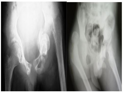
In many regions of the developing world, pediatric osteomyelitis and septic arthritis are still a major clinical and economic burden, with the incidence of these conditions being much higher in comparison with the developed world [1,2,3]. Early detection and prompt initiation of management are integral to avoid potential serious and permanent complications [4]. Several factors, both cultural and economic, lead to misdiagnosis and/or under-treatment of these children in low-resource settings and, as a consequence, patients present with severe complications. For example, in major joints such as the hip, the infection can lead to subluxation, dislocation and fracture-dislocation (Figure 1). When fracture-separation happens the necrotic head remains in the acetabular cavity however, very rarely, it can migrate inside the pelvis. We report two cases of this rare and unusual clinical course in two children who presented to Comprehensive Rehabilitation Services in Uganda (CoRSU) Hospital.

Figure 1: Radiographs of a 12-year (left) and 14-year-old boy (right), showing the variety of presentations in untreated septic arthritis. Usually the acetabular floor prevents the intra-pelvic migration of the head, even in presence of pathological fracture or severe infection.
Case 1
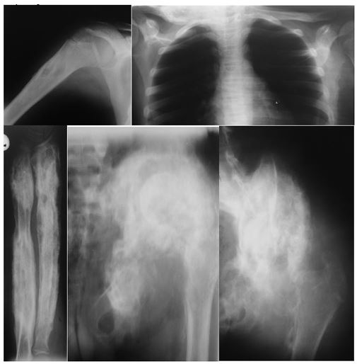
A fourteen-year-old boy was brought to our facility with several months’ history of multiple sites of osteo-articular infections. He had previously been treated conservatively with antibiotics. At presentation he was ill-looking, pale and malnourished. He mobilized for short distances only, with a limp and with the aid of a long stick. On examination he had a discharging sinus in each of the following locations: right arm, left forearm, left thigh, left trochanteric area and over the right sterno-clavicular joint. In addition, he had a soft mass, resembling a cold abscess, along the lateral border of the left scapula. On admission all the affected sites were x-rayed (Figure 2) and the working diagnosis was of disseminated osteo-articular tuberculosis. The boy was started on a hyper-caloric diet and transfused whist awaiting surgery.
The first surgery was done roughly two months after the admission; a concurrent debridement of the left hip, right clavicle and left radius and ulna was carried out. An abscess in the mid-third of the left thigh was incised as well. Biopsy results were negative for tuberculosis. Six months later a further surgery for hip debridement was done but the necrotic femoral head, well visible on the x-ray film, was not found. A retained gauze, most probably used in the previous dressing change and located in the subcutaneous space, was also removed. Due to persistent drainage from the left hip, a further six months later, further hip debridement was done, but again the head was not found. A minor debridement of the radius was also done in the same operating session and new specimens were taken for histology. These biopsies confirmed our suspicion of osteoarticular tuberculosis. At this stage, he was started on a 12 months long anti-tuberculous treatment. As expected his general condition improved rapidly; all the previously affected sites healed except for the persistent sinus in the hip area. Once again a debridement of the left ilium was done and biopsy was taken. It resulted negative for TB. The gluteal region was widely explored but no femoral head was found.
With the necrotic head still in-situ a new surgery was planned. This time the incision was done along the iliac crest and, finally, the femoral head was found lying flush to the inner side of the ilium and covered by a thin bony roof. The head was enclosed in a kind of osseous box. After its removal, there was a dramatic improvement of the local conditions. More than 16 months later the boy re-presented complaining of a slight discharge from the hip area, over the iliac crest. A minor debridement was done and the biopsy was repeated. It resulted negative for tuberculous infection. The boy was followed-up for two years after the last debridement, after which he was unfortunately lost to follow-up for 10 years. He was traced again in 2022. He is now a 29-year-old man, married, and a farmer by profession. He appeared in good general conditions with no signs of infection relapse. He was independent with his activities of daily living and mobilized without the need for assistive devices

Figure 2: Radiographs showing disseminated osteo-articular tuberculosis on admission. The loss of normal architecture and texture of the left hemipelvis prevented a prompt diagnosis of intrapelvic location of the necrotic head.
Case 2
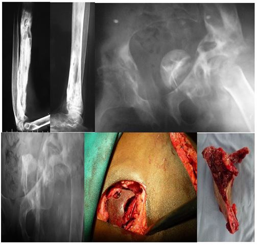
A 14-years-old girl presented to our facility with a two-year history of multiple and recurrent sites of osteo-articular infections. She presented after several months of hospitalization in different remote health facilities where conservative treatment was given. At presentation the girl was ill-looking, pale, malnourished and was carried in by her mother, as she was unable to stand. She had been unable to walk for the past two years due to flexion contractures of the hips and knees. Sinuses were detected in the left forearm, right leg and in the perineal area, just lateral to the left labium major. An extensive dermatitis was also present. Her ESR was 94 mm/hour and the hemoglobin was 6.9 g/dl. All the affected sites were x-rayed (Figure 3) and a staged surgical plan was proposed. The girl started a nutritional rehabilitation plan and was transfused as necessary.
The first surgery was directed at the debridement of the right tibia and the left radius, in association with manipulation of the knees in extension. A few weeks later a debridement of the hip area was done and specimens for biopsies were taken. They resulted negative for tuberculosis.

Figure 3: Radiographs on admission showing the extensive osteomyelitis of the radius, the cystic type infection of the tibia and the location of the necrotic head in the lower pelvis. The intraoperative position of the head, flush to the bladder, and the retrieved fragment of acetabular floor are shown with the corresponding radiograms.

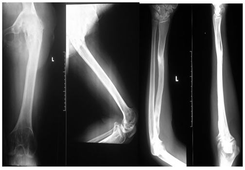
Figure 4: Radiographs taken at the last control, five years later. Spontaneous hip fusion, without signs of infection relapse, and complete remodeling of the radius.
The first surgery was directed at the debridement of the right tibia and the left radius, in association with manipulation of the knees in extension. A few weeks later a debridement of the hip area was done and specimens for biopsies were taken. They resulted negative for tuberculosis.
Ten days later a new hip debridement was coupled with a bikini incision in the left side of the lower abdomen and the necrotic femoral head was found lying flush to the wall of the bladder. The post-operative X-ray demonstrated that a necrotic acetabular floor fragment was left in-situ the abdominal cavity; unfortunately, no fluoroscopic control was done in theater. The fragment was removed a few days later.
The girl’s condition improved and she was discharge home, able to walk with a single crutch and supported with knee braces.
Four months later, she re-presented complaining of moderate discharge in the hip area, over the greater trochanter, and in the left forearm. Surgery for concurrent debridement of the two sites was carried out and healing was achieved in a couple of weeks. At the last follow up, now aged 18 years, she presented in good general conditions with no infection relapse. The left hip fused spontaneously at 60 degrees of flexion and 10 degrees of abduction while the knee had a normal ROM. Radiographs of the hip and forearm were taken (Figure 4). Importantly she didn’t report any pain, she was able to walk without aids and she was able to perform all the activities of daily living independently. Advice was given for an operation of valgus-extension sub-trochanteric osteotomy to improve her gait and function.
Discussion
Septic arthritis of major joints, namely hip and knee, preferentially affects children living in low and middle income countries. Early detection and prompt treatment are a must for successful management and for prevention of severe complications. Sadly, these goals are rarely achieved in countries coping with fragile and underfunded health systems. It is, therefore, unsurprising that children continue to present to our facility with the sequelae of neglected or undertreated infections. Both patients from the above case reports were from families below the poverty line, obliged to make a living with 2 USD per day. Quite often, what seems to a surgeon a kind of neglect of severely ill children is simply a side-effect of poverty and the associated poor living conditions.
In long standing cases diagnosis is easily reached on clinical and radiographic grounds. Pain, limb deformity, loss of joint function, discharging sinuses are rarely missed on clinical examination. Plain radiograms are taken to confirm the diagnosis. Further imaging is usually not necessary; however, a CT scan would have been helpful in the first case report by reducing the number of hip debridement surgeries performed. Unfortunately, CT imaging is not available in our setting.
In disseminated cases, particularly in countries where the infection is endemic, tuberculosis should always be considered a differential diagnosis. Intraoperative biopsy is, therefore, mandatory and may need to be repeated, especially if the results are in contrast with the clinical suspicion as highlighted in the first case.
The clinical documentation of the two cases showed that antibiotics were widely used prior to presentation to our facility. It is known that they are of dubious benefit in these chronic conditions and unfortunately, a considerable amount of money was spent by these families on this treatment strategy.
Optimization of the patients before surgery mainly relies on nutritional rehabilitation and blood transfusions. Psychological support and early return to recreational activities were significant in restoring hope and trust in the children’s relatives. Disseminated cases require multiple surgeries making the cost of treatment hardly affordable to the families. Since there is no coverage from the underfunded national health system these children might be considered, to a certain extent, hostages of the struggling economies in which they are living. Fortunately, these two cases where funded by humanitarian help.
Most cases are still seen in peripheral health units, where urgent medical attention to a febrile and crying child is lacking. At the moment, it is difficult to imagine a better scenario in the coming years in Uganda, taking into account the growing pediatric population and the fragile economy. It is sad to think that many children will likely continue to suffer from preventable consequences of mistaken diagnoses and lack of urgent treatment.
References
- Chiappini E, Mastrolia MV, Galli L, De Martino M, Lazzeri S. Septic arthritis in children in resource limited and non-resource limited countries: an update on diagnosis and treatment. Expert Rev Anti Infect Ther. 2016; 14: 1087-1096.
- Omoke NI. Childhood pyogenic osteomyelitis in Abakaliki, south east Nigeria. Niger J Surg. 2018; 24: 27-33.
- Lavy CB. Septic arthritis in Western and sub-Saharan African children-a review. Int Orthop. 2007; 31: 137-44.
- Tamer E, Shady M. Acute osteoarticular infections in children are frequently forgotten multidiscipline emergencies: beyond the technical skills. EFORT Open Rev. 2021; 6: 584-592.

