Case report - Volume 3 - Issue 3
Osteoma of external auditory canal: A case report
Dr. Shriya Gupta1*; Dr. Aditya Yeolekar2; Dr. Shivani Mishra3; Dr. Manodnya Jalvi3
1Senior resident, Department of Otorhinolaryngology, Postgraduate Institute and Yashwantrao Chavan Memorial Hospital, Pimpri, Maharashtra, India.
2Associate Professor, Department of Otorhinolaryngology, Postgraduate Institute and Yashwantrao Chavan Memorial Hospital, Pimpri, Maharashtra, India.
3Junior resident Department of Otorhinolaryngology, Postgraduate Institute and Yashwantrao Chavan Memorial Hospital, Pimpri, Maharashtra, India.
Received Date : April 07, 2023
Accepted Date : May 11, 2023
Published Date: May 18, 2023
Copyright:© Shriya Gupta 2023
*Corresponding Author : Dr. Shriya Gupta, Senior resident, Department of Otorhinolaryngology, Postgraduate Institute and Yashwantrao Chavan Memorial Hospital, Pimpri, Maharashtra, India.
Email: drshriya29@gmail.com
DOI: Doi.org/10.55920/2771-019X/1441
Abstract
Osteomas are benign bone neoplasms most commonly found in the frontoethmoidal area. Temporal bone osteoma is a rare entity. External auditory canal osteoma present as a solitary, unilateral and slow-growing pedunculated mass in the bony canal. Here, we present an unusual presentation of this entity.
Keywords: External auditory canal, osteoma, surgical excision
Case report
A 36-year-old female patient presented with a gradual decrease in hearing in the right ear since 1 year and dull aching pain since 2 months. It was associated with itching, chronic irritation and cerumen impaction in the ear. It was associated with itching, chronic irritation and cerumen impaction in the ear. No history of tinnitus or vertigo. On examination, a hard, non-tender mass covered with skin completely occluding the external auditory canal was seen. (Figure 1A) Probing around the mass showed posterosuperior and anteroinferior attachment. Pure tone audiometry revealed moderate conductive hearing loss (40 dB) in the right ear at hearing frequency ( Figure 3A) with normal hearing in the left ear.

Figure 1: A)Endoscopic examination of the right ear revealed a hard mass completely occluding canal with normal skin. Figure 1B)High resolution computed tomography showing coronal view of both temporal bone. Right sided, red circle represent bony lesion arising from the tympanosquamous suture line. Figure 1C) High resolution computed tomography showing axial view of both temporal bone. Right side external auditory canal showing irregular , broad based mass arising from the posterior wall of canal marked with yellow circle. Tympanic membrane is visualised separately from the lesion

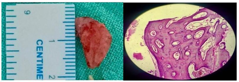
Figure 2: A)Gross specimen of the external auditory canal; Figure 2B) Histopathological specimen showing fibrovascular channels with bony lamellae suggestive of osteoid osteoma.

Figure 3: A)Pure tone audiogram of the patient showing moderate conductive hearing loss preoperatively. B) Pure tone audiogram of the patient showing normal hearing postoperatively. Figure 3C) Endoscopic view of the canal and tympanic membrane at 1 month follow up.
High resolution computed tomography of both the temporal bone showed 2 1 cm2 sized ovoid, computed tomography of both the temporal bone showed 2 1 cm2 sized ovoid, hyper-dense, pedunculated broad based bony mass completely occluding external auditory canal arising from tympanosquamous and tympanomastoid suture line with medial displacement of tympanic membrane. The rest of the mastoid cavity and ossicles were normal (Figure 1B,1C). A provisional diagnosis of right external auditory canal osteoma was made. Transcanal excision of the mass was planned.
A 2mm osteotome was used to break the posterior and superior attachments of the mass following which it was removed in toto. After the removal of the mass , epithelial debris was visualised and suctioned. Attic region and pars tensa was normal. This excised ovoid mass was sent for histopathological reporting. Irregular bony canal was saucerised using diamond burr and wide canaloplasty was done. The bare area of canal was covered with temporalis fascia. External auditory canal was packed with antibiotic soaked gel foam. A betadine soaked merocele was kept in canal which was removed after 10 days.
Gross specimen showed 1.5 cm grey-white mass, which was hard on palpation, gritty to cut (Figure 2A). Histopathological reporting suggested of osteoid osteoma which consisted of fibrovascular channels covered with periosteum and squamous epithelium with lamellated bone and minimal osteocytes (Figure 2B).
Postoperatively, follow up was done at 1 month which showed normal hearing in both the ears with healed wide canal (Figure 3).
Discussion
Osteomas are benign bone neoplasms most commonly found in the frontoethmoidal area. Temporal bone osteoma is a rare entity [1]. They are seen in squamous, mastoid, internal and external auditory canal, glenoid fossa, middle ear, eustachian tube, petrous apex and styloid process. External auditory canal osteoma present as a solitary, unilateral and slow-growing pedunculated mass in the bony canal [2]. It’s incidence peaks in the fourth decade of life, and a male-to-female ratio is known to be 2-3:1. These are tumours consisting of mature lamellar bone. These are very well documented in the external auditory canal but they are otherwise very rare in the temporal bone. The most common site of origin appears to be the mastoid with about 20% developing in the middle ear. Treatment of osteoma is surgical removal through its pedicle to avoid recurrences.
Exostoses or Osteomas are generally considered as differential diagnosis for bony outgrowths in the external ear. The diagnosis is based on combination of clinical history and examination, radiological imaging and histopathology. Most widely accepted theory of origin for exostosis is a reaction to cold-water stimulation of the local periosteum, while osteomas are benign osseous tumours. Histologically, osteomas have internal fibrovascular channels surrounded by irregularly oriented lamellated bone and minimal osteocytes. Graham2 described exostoses as broad-based, dense and composed of lamellated bone, covered with periosteum and its overlying squamous epithelium with an internal structure formed by concentric, dense layers of subperiosteal bone with abundant osteocytes. Radiologically, external auditory canal osteomas usually appear as solitary, pedunculated, well-circumscribed lesions with high attenuation, equivalent to that of bone density. Exostoses are generally multiple, bilateral, symmetrical bony elevations attached to the canal with a broad base and lie close to the tympanic membrane. Computed tomography is also usually useful for defining the association between lesion size and the surrounding tissue [3,4]. Therefore, preoperative imaging is valuable for the differential diagnosis of osteoma and exostosis.
Osteomas in the external auditory canal do not require treatment till symptoms such as conductive hearing loss, recurrent infection secondary to cerumen impaction or a cholesteatoma develop. They are typically asymptomatic and can be monitored. The size and location of the osteoma determines the surgical approach.
Sheehy et al.5 reported that external auditory canal osteomas could be surgically removed through two microscopic approaches depending on their location related to the isthmus of external auditory canal. When external auditory canal osteomas originated lateral to the isthmus, they could be removed using a transmeatal approach. However, osteomas medial to the isthmus required postauricular approach. Grinblat at al [6] also proposed two surgical approaches in canaloplasty for exostosis and osteoma of the external auditory canal depending on the size of the lesion. A simple transcanal approach was applicable if the size was less than 50% stenosis, whereas a retroauricular transcanal approach was advised if the stenosis was greater than 50%.
Conclusion
Osteomas are benign bony neoplasms that show a predilection for the external auditory canal. However, these lesions arising from temporal bones are considered rare [7]. report their incidence to be 0.05% of complete otologic surgery.8 Osteoma are mostly asymptomatic but the lack of epidermal clearance and aeration are followed by complaints of hearing loss to local infection and otorrhea. Surgical excision is indicated only if patient becomes symptomatic. Local monitoring and follow up can decrease the recurrence of the disease.
References
- Burton DM, Gonzalez C. Mastoid osteomas. Ear Nose Throat J. 1991; 70: 161–2.
- Graham MD. Osteomas and exostoses of the external auditory canal. A clinical, histopathologic and scanning electron microscopic study. Ann Otol Rhinol Laryngol. 1979; 88(4 Pt 1): 566-572.
- Ebelhar AJ, Gadre AK. Osteoma of the external auditory canal. Ear Nose Throat J. 2012; 91(3): 96-100.
- Brea B, Fidalgo AR. Imaging diagnosis of benign lesions of the external auditory canal. Acta Otorrinolaringologica (English Edition). 2013; 64(1): 6-11.
- Sheehy JL. Diffuse exostoses and osteomata of the external auditory canal: a report of 100 operations. Otolaryngology--head and neck surgery: official journal of American Academy of Otolaryngology-Head and Neck Surgery. 1982; 90(3 Pt 1): 337-42.
- Grinblat G, Prasad SC, Piras G, He J, Taibah A, Russo A, Sanna M. Outcomes of drill canalplasty in exostoses and osteoma: analysis of 256 cases and literature review. Otology & Neurotology. 2016; 37(10): 1565-72.
- Yamasoba T, Harada T, Okuno T, et al. Osteoma of the middle ear. Report of a case. Arch Otolaryngol Head Neck Surg. 1990; 116: 1214-6.
- Shankar Shah ST, Chetri S, Manandhar A, Pokhrel B, Aryal A. Prakash. Osteoma of the External Auditory Canal Masquerading as an Aural Polyp: Case Report and Review of Literature. American Journal of Medical Case Reports. 2013; 1(1): 3-5.

