Case report - Volume 3 - Issue 3
Angiosarcoma of the scalp : A zosteriform presentation
Hajar Tahiri1*; Fouzia Hali1; Farida Mernissi2; Soumaya Chiheb1
1Dermatology department, UHC Ibn- Rochd, Casablanca, Morocco.
2Anatomical pathology, UHC Ibn- Rochd, Casablanca, Morocco.
Received Date : April 16, 2023
Accepted Date : May 23, 2023
Published Date: May 30, 2023
Copyright:© Hajar Tahiri 2023
*Corresponding Author : Hajar Tahiri, Dermatology department, UHC Ibn- Rochd, Casablanca, Morocco.
Email: Tahirihajar33@gmail.com
DOI: Doi.org/10.55920/2771-019X/1449
Abstract
Cutaneous angiosarcoma is a rare and agressive tumor with poor outcome, and early diagnosis is paramount, but its often delayed because of the long delay in the appearance of lesions and the variable clinical presentation of this neoplasia. This article reports the case of an 84-year-old patient who presented with persistant ulcerated placard of the scalp and face evolving since one year and treated as herpes zoster ophthalmicus. Incisional biopsy revealed a morphological aspect of high grade angiosarcoma with CD 31 + / CD 34 -. An extension workup made of thoraco-abdominal and pelvic CT scans showed hepatic, adrenal and bone metastasis. Given his advanced age, and his general condition and visceral metastasis at the moment of diagnosis, the decision of the multidisciplinary concertation meeting was to put him under palliative care, he passed away 8 months after the diagnosis of angiosarcoma was made, 18 months after the occurrence of the first symptoms. Cutaneous angiosarcoma (CA) is a rare aggressive malignant tumor, that occurs in elderly white man. Because of the variability of its presentation and its insidious evolution, the appearance of lesions may not concern patients or practitioners for some time,Therefore correct diagnosis may be delayed, often dramatically limiting the tools of treatment options and altering the outcome of the patients.
Keywords: Ulcerated placard, Cutaneous angiosarcoma, neoplasm, Scalp, herpes zoster ophtalmicus
Introduction
Cutaneous Angiosarcoma is an aggressive malignant tumor of the vascular endothelium that accounts for approximately 1,6 % of cutaneous soft tissue sarcomas [1]. It mostly occurs in elderly white men, and can appear anywhere on the skin with a predilection for head and neck in 60% of cases and can be unifocal or multifocal at presentation [2]. Timely diagnosis is often delayed, because of the very variable clinical presentation of this tumor and its slow onset and thus a dark prognosis with a 5-year overall survival less than 26% [1]. We report the case of a patient with Angiosarcoma of the scalp mimicking an herpes zoster ophthalmicus.
Observation
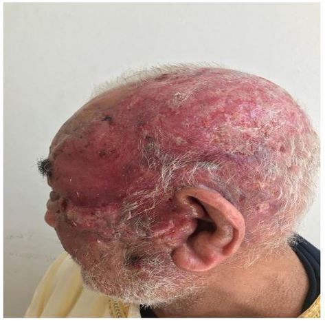
An 84 year man, farmer with a history of diabetic and arterial hypertension for 20 years, was admitted to our institution for indurated and ulcerated purple plaques on the scalp that had been evolving one year prior to consultation and treated as an herpes zoster ophthalmicus on several occasions. General clinical examination revealed a conscious patient, with a ECOG Scale of Performance Status : 2, dermatological examination found an ulcerated and indurated purplish scalp plaques, which seemed to occupy the territory of the left ophthalmic nerve with a circumferential extension to the right hemiscalp [figure 1]. This plaques extended to the forehead and left cheek, with multiple ulcerated nodules, we also found edema of both eyelids more pronounced on the left eye, obscuring the vision [figure 2]. Further examination revealed multiple macroscopique tender lymph nodes occupying the left cervical region. An incisional biopsy was performed and revealed a morphological aspect of high grade Angiosarcoma with CD 31 + / CD 34 -. An extension workup made of thoraco-abdominal and pelvic CT scans showed hepatic, adrenal and bone metastasis. The patient was discussed in a multidisciplinary concertation meeting and the decision was to put him under palliative care, given his advanced age, general condition and visceral metastasis. He passed away eight month later, about 18 months after the occurrence of the first symptoms of the tumor.

Figure 1: Ulcerated and indurated purplish scalp plaques.
Discussion
The originality of our observation lies in the atypical aspect of the lesions, simulating an herpes zoster ophtalmicus. Angiosarcoma is a rare and very aggressive tumor that develops in the vascular endothelial cells of the skin and superficial soft tissues, it mostly occurs in elderly people, men are more affected than women, the main risk factors are ionizing radiation and the longstanding solar exposure [3], as was the case of our patient. Reported cases in the literature have described the lesions as infiltrated purplish-red papules, plaques, and nodules that may be misdiagnosed as rosacea, eczema, and hematoma. These lesions may also mimic cutaneous lymphoma, metastatic carcinoma or Kaposi's sarcoma [3]. To our knowledge, this is the first case of Angiosarcoma taking on a metameric arrangement and thus the diagnosis of herpes zoster ophtalmicus complicated by cellulitis and treated with antivirals and antibiotics was given before the correct diagnosis of Angiosarcoma provided.
As in our patient's case, 50% of Cutaneous Angiosarcomas occur in the head and neck. The scalp is the most affected area, which also holds the worst prognosis, The 5-year survival rate for patients with Cutaneous Angiosarcoma of the head and neck ranges from 10% to 54% [2].
Prompt diagnosis and treatment is an important factors in improving survival, but may be delayed due to insidious tumor progression as lesions can be hidden by hair for a long time and the initial benign presentation, as was the case for our patient. Jonathan M.et Al showed that the average delay between the appearance of the lesions and the positive diagnosis was 4 months, in our case the delay was one year [2]. Despite its infiltrative nature, visceral metastases of Angiosarcoma are uncommon. In a study of 52 patients, only 2% had distant metastases [2]. Their presence at the time of diagnosis is considered as a poor prognostic factor, as was the case of our patient.
Several treatments are available for Angiosarcoma. Traditionally, the combination of a large surgical excision the tumor and preoperative or postoperative radiotherapy provides the basic treatment [4]. However, complete surgical resection can be challenging because of the very infiltrative and multifocal nature of the tumor like was the case of our patient, which often leads to local recurrence and metastatic disease
The median survival time of patients with Cutaneous Angiosarcoma of the scalp ranges from 12 to 42 months [5]. the median survival of our patient since the first appearance of the lesions to the time of diagnosis was 18 months. Age over 70 years, tumor size larger than 5 cm, location in the scalp , multifocality ,ulceration and metastases in the moment of diagnosis are predisposing factors for poor prognosis of Angiosarcoma, our patient had them all.
Conclusion
Autaneous Angiosarcoma is an aggressive malignant tumor, its variable presentation and insidious oncet of the lesions may not trigger concern among patients or practitioners for some time and thus correct diagnosis can be delayed, often drastically altering outcome and treatment options.
Our case serves as a reminder to physicians that an abnormal skin finding in elderly adults should prompt them to suspect Angiosarcoma and perform an early biopsy.
References
- Wilssens NO, Den Hondt M, Duponselle J, Sciot R, Hompes D, Nevens THG. An uncommon presentation of a cutaneous angiosarcoma. Acta Chir Belg. 2021; 121(5): 351‑3.
- Bernstein JM, Irish JC, Brown DH, Goldstein D, Chung P, Razak ARA, et al. Survival outcomes for cutaneous angiosarcoma of the scalp versus face. Head Neck. 2017; 39(6): 1205‑11.
- Ronchi A, Cozzolino I, Zito Marino F, De Chiara A, Argenziano G, Moscarella E, et al. Primary and secondary cutaneous angiosarcoma: Distinctive clinical, pathological and molecular features. Ann Diagn Pathol. 2020; 48: 151597.
- Florou V, Wilky BA. Current and future directions for angiosarcoma therapy. Curr Treat Options Oncol. 2018; 19(3): 14.
- Guadagnolo BA, Zagars GK, Araujo D, et al. Outcomes after definitive treatment for cutaneous angiosarcoma of the face and scalp. Head Neck. 2011; 33(5): 661-667

