Clinical Image - Volume 3 - Issue 3
Newborn with epispadias
Jesus Ruiz*
Department of Family Medicine, University of North Carolina, Chapel Hill, North Carolina, USA.
Received Date : April 20, 2023
Accepted Date : May 26, 2023
Published Date: June 02, 2023
Copyright:© Jesus Ruiz 2023
*Corresponding Author : Jesus Ruiz, Department of Family Medicine, University of North Carolina, Chapel Hill, North Carolina, USA.
Email: jesus_ruiz@med.unc.edu
DOI: Doi.org/10.55920/2771-019X/1452
Clinical Image
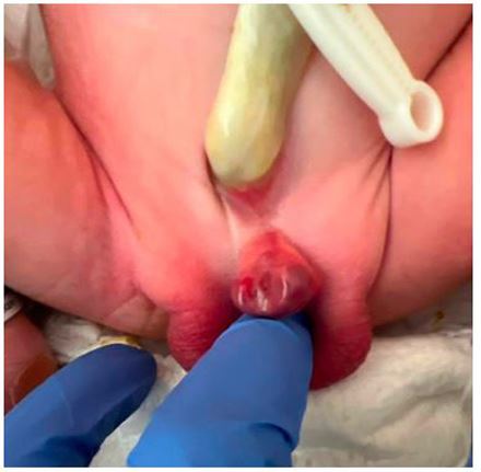
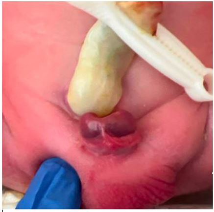
A newborn was examined. The patient was born to 29-yearold women at 38 weeks and 5 days via normal spontaneous vaginal delivery. Prenatal care was uncomplicated. Fetal anatomy ultrasound and labs were within normal limits. Vital signs were stable and physical exam revealed atypical genitalia (Figure 1 and 2). Urinary incontinence with palpation of the abdomen was noted. Given the findings of absence of the dorsal aspect of the urethra and overlying skin we made the diagnosis of epispadias. Diagnosis is based on clinical and physical exam findings during routine newborn examination. Typical exam findings in males include short phallus, dorsal meatus, dorsal chordee, splaying of the corpora cavernosa and ventral foreskin [1]. Epispadias is a rare urogenital malformation resulting from failure of the urethral tube to tabularize on the dorsal aspect. The exact etiology of epispadias remains unknown however it is believed to be related to cloacal membrane development [2]. Isolated epispadias is rare with an incidence of 1 per 120,000 in males and 1 in 500,000 females [3]. The differential diagnosis for abnormal male genitalia includes classic bladder exstrophy, cloacal exstrophy, and hypospadias. A plain radiograph can be obtained to document pubic diastasis and an abdominal ultrasound to rule out associated congenital anomalies of the upper urinary tract, especially in patients with incontinent epispadias [1]. Treatment of epispadias is surgical. The aim of surgery is to reconstruct the urethra, close the defect, and provide optimal function and cosmetic results.

Figure 1: Epispadias. Dorsal meatus, dorsal chordee, splaying of the corpora cavernosa and ventral foreskin.

Figure 2: Epispadias. Short separation between penis and the umbilical stump.
References
- Grady RW, Mitchell ME. Management of epispadias. Urol Clin North Am. 2002 May;29(2):349-60, vi.
- Anand S, Lotfollahzadeh S. Epispadias. [Updated 2022 Dec 3]. In: StatPearls [Internet]. Treasure Island (FL): StatPearls Publishing.
- Hall SA, Manyevitch R, Mistry PK, Wu W, Gearhart JP. New Insights on the Basic Science of Bladder Exstrophy-epispadias Complex. Urology. 2021; 147: 256-263.

