Clinical Image - Volume 3 - Issue 3
Dermoscopy of lipoid necrobiosis
Sabrina Oujdi*; Meryem Soughi; Siham Boularbah; Hanane Baybay; Sara Elloudi; Zakia Douhi; Fatima Zahra Mernissi
Department of Dermatology, University Hospital Hassan II, Fes, Morocco.
Received Date : May 05, 2023
Accepted Date : June 02, 2023
Published Date: June 09, 2023
Copyright:© Oujdi Sabrina 2023
*Corresponding Author : Oujdi Sabrina, Department of Dermatology, University Hospital Hassan II, Fes, Morocco
Email: sabrinaoujdi92@gmail.com
DOI: Doi.org/10.55920/2771-019X/1458
Clinical Image
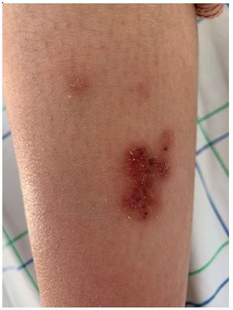
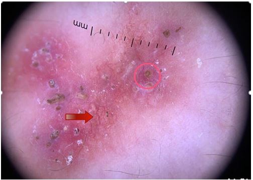
Lipoid necrobiosis is a rare granulomatous dermatosis, it usually occurs in diabetic subjects. Clinically, it presents as papules, nodules or erythematous plaques with a yellowish atrophic center, sometimes telangiectatic, and is located preferably at the pre-shin level. the confirmation of the diagnosis is based on the histology spectra which will find a granulomatous infiltrate arranged in palisades occupying the whole height of the dermis with an infiltrate composed of lymphocytes, histiocytes, plasma cells and epithelioid cells, as well as epithelioid cells and giant cells. Vascular lesions can be observed [1]. Few publications in the literature have described the dermoscopic aspect of lipoid necrobiosis which has a yellow-orange background testifying to the presence of granulomas and the presence of clear serpiginous vessels, due to the epidermal atrophy that accompanies the pathology, long and branched vessels like lightning bolts zigzagging the sky [2]. We describe a new case of dermoscopy of lipoid necrobiosis. A 18-year-old woman diabetic under insulin for 6 years, reports for 1 year an erythematous papule on left leg progressively increasing in size and asymptomatic for which she applied by self-medication topical antibiotics without improvement. Clinical examination found an erythematous squamous-crusty plaque with irregular contours on the anterior face of the lower left leg. Dermoscopy revealed a diffuse yellow-orange background and clear arborescent vessels , scales and keratin plugs.Histopathologic study was compatible with lipoid necrobiosis.
Consent: The examination of the patient was conducted according to the Declaration of Helsinki principles.
Conflicts of interest: The authors do not declare any conflict of interest

Figure 1: Clinical picture showing an erythematous plaque.

Figure 2: Dermoscopic picture showing: diffuse yellow-orange background , clear arborescent vessels (red arrow). keratin plugs (red circle).
References
- L’image du mois Nécrobiose lipoïdique G, Szepetiuk C, Piérard-Franchimont M-A, Reginster G.E, Piérard. Rev Med Liège. 2011; 66 : 2 : 61-63
- Razmi TM, Sawatkar GU, Sekar A, Vinay K, De D, Saikia UN, Parsad D. Dermoscopy of palisaded neutrophilic and granulomatous dermatitis. Clinical and Experimental Dermatology. 2019.

