Clinical Image - Volume 3 - Issue 3
Localized hypopigmentation under the Dermoscope: A diagnostic conundrum
Kenza Tahri Joutei Hassani*; Zakia Douhi,Hanane Baybay; Sara Elloudi,Meryem Soughi; Fatima Zahra Mernissi
Department of Dermatology, University Hospital Hassan II, Morocco.
Received Date : May 10, 2023
Accepted Date : June 22, 2023
Published Date: June 29, 2023
Copyright:© Tahri Joutei Hassani Kenza 2023
*Corresponding Author : Tahri Joutei Hassani Kenza, Department of Dermatology, University Hospital Hassan II,Fes, Morocco.
Email: kenzatahri10@gmail.com
DOI: Doi.org/10.55920/2771-019X/1471
Clinical Image
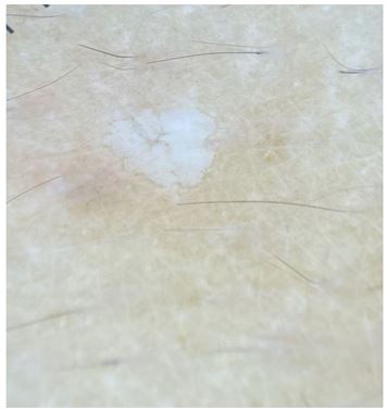
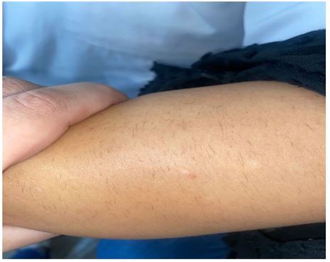
Patients with hypopigmented skin conditions may face cosmetic and psychological difficulties due to the significant difference in appearance between the affected skin and the surrounding normal skin. As a result, they may feel the need to seek evaluation and treatment [1]. On occasion, we have encountered patients with acquired well- demarcated, scattered hypopigmented papules which can suggest multiples diagnosis such as warts, guttate hypomelanosis or an hypopigmented variant of seborrheic keratosis.Seborrheic keratoses are a common type of acquired skin lesion in adults. While they typically appear as brown or black macules or papules, they can rarely presented with a pale, hypopigmented papule with a surface that displayes a variation in color [2]. A notable clinical feature that sets seborrheic keratoses apart is their "stuck on" appearance. This means that the lesion's edges are palpable and differ from the surrounding skin, giving the impression that it was affixed to the skin [2].We report the case of a 30 year old woman presenting a white papule on the right forearm. Clinical examination revealed a slightly infiltrated whitish 3 mm papule suggesting a wart, a hypopigmented seborrheic keratosis or a hyperkeratotic guttate hypomelanosis (Figure1). Dermoscopy showed a good demarcation in the periphery with a bitten aspect and some pseudocysts (Figure 2). A biopsy was performed confirming the diagnosis of seborrheic keratosis.

Figure 1: Slightly infiltrated white papule on the right forearm.

Figure 2: Dermoscopy showing a good demarcation in the periphery with a bitten aspect and some pseudocysts.
References
- Poon S, Beach RA. Localised hypopigmentation: clarification of a diagnostic conundrum. Br J Gen Pract. 2018.
- Kim SK, Park JY, Hann SK, Kim YC, Lee ES, Kang HY. Hypopigmented keratosis: is it a hyperkeratotic variant of idiopathic guttate hypomelanosis? Clin Exp Dermatol. 2013.

