Case report - Volume 3 - Issue 4
Epithelioid hemangioendothelioma of the brachial artery: A rare benign vascular tumor
Enes Ozsozgun; Esin Kurtulus Ozturk*; Saffet Ozturk
Department of Radiology, Ankara Etlik City Hospital, 06170, Turkey.
Received Date : May 25, 2023
Accepted Date : July 06, 2023
Published Date: July 13, 2023
Copyright:© Esin Kurtulus Ozturk 2023
*Corresponding Author : Esin Kurtulus Ozturk, Department of Radiology, Ankara Etlik City Hospital, 06170, Turkey.
Email: e.kurtulus@hotmail.com
DOI: Doi.org/10.55920/2771-019X/1490
Introduction
Epithelioid hemangioendotheliomas are uncommon vascular tumors presenting with different biological behavior and clinical features. It is a pathology that should be diagnosed early because it is seen at earlier ages and can cause distant organ metastases. Due to its rarity, it is often not included in the differential diagnosis of soft tissue masses [1]. In this case, we aimed to present a malignant epithelioid hemangioendothelioma presenting with palpable swelling.
Case presentation
A 75-year-old male patient was admitted to the orthopedics clinic due to swelling on the medial side of his right arm. On ultrasonography, a 32x20x25mm ill-defined, lobulated soft tissue mass surrounding the brachial artery located medial to the right arm was observed.Significant peripheral vascularization was noted on Doppler examination (Figure 1).
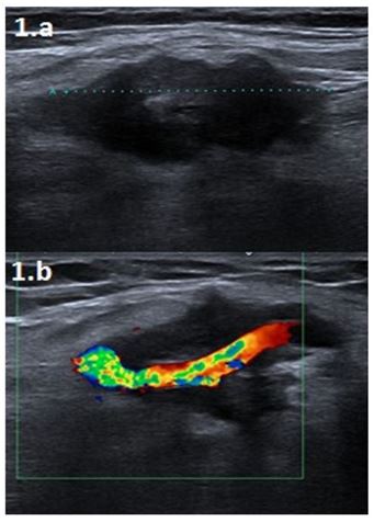
Figure 1: B-mod and colour Doppler ultrasound images (a and b) show an irregularly contoured soft tissue mass surrounding the brachial artery medially in the right arm midsection.
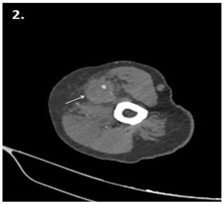
Figure 2: Axial upper extremity CT angio image shows irregular course of the brachial artery within the mass (arrow).
Irregular course of the brachial artery in the mass was observed on upper extremıty Ct angio (Figure 2). The magnetic resonance imaging scan revealed a heterogeneous mass with irregular contours, surrounding the brachial artery and indistinguishable from the brachial vein (thrombosis), restricting diffusion, and showing peripheral irregular contrast enhancement was observed (Figure 3). The imaging findings suggested a vascular tumor but with difficulty in differentiating malignant or benign. After surgical excision of the lesion, the histopathology was reported as malignant epithelioid hemangioendothelioma originating from the brachial artery.
Discussion
Epithelioid hemangioendothelioma was first described in 1982 and was previously known as intravascular bronchioalveolar tumor. Histologically, it consists of pleomorphic spindle epithelioid cells with high mitotic activity. It contains various immunohistochemical endothelial markers (CD31, CD34, Fli-1 etc.). In its etiopathogenesis, Some molecules and chromosomal mutations are thought to cause the stimulation of angiogenesis and endothelial cell proliferation [1].
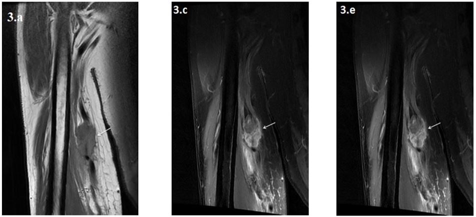
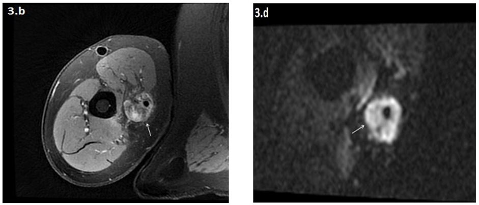
Figure 3: Axial T1W (a), fat-sat axial (b) and coronal (c) PD images show a heterogeneous mass lesion (arrow) with irregular contours surrounding the brachial artery, but where the brachial vein cannot be distinguished (thrombosed). Diffusion (d) and contrast-enhanced coronal T1W(e) images show that the lesion is diffusion-restricted and peripherally irregularly enhanced.
Epithelioid hemangioendotheliomas are rare tumors of vascular origin that usually occur in young adults. In the literature, there are disagreements about the incidence of the most frequently involved organ and tumor. Epelboym et al. stated that the most frequently involved organs were liver, lung and bone, respectively, while in the study of Requena and Kutzner, more than half of the cases were related to vascular structures [2,3]. Epithelioid hemangioendotheliomas are mostly seen in the vascular structures of the venous system (most commonly the femoral vein) [4].
More rarely, it presents as a painful mass lesion originating from arterial vascular structures. It can be confused with aneurysms and arteriovenous malformations as it wraps around the vascular structures in the form of a sheath [5]. Depending on the tumor's occlusion of the vascular structure, findings such as edema and thrombophlebitis may be observed. Distant organ metastases are most common to the lung [4,6]. Fewer cases were reported in the upper extremity arteries than in the lower extremity arteries. Cases of epithelioid hemangioendothelioma seen in the superficial palmar arch, ulnar artery and radial artery have been reported in the literature [7-9].
On ultrasound, it is observed as a hypoechoic mass surrounding the vascular structure to a certain extent, adjacent to the vascular structure. In Doppler ultrasound examination, it is in the form of a mass with vascular blood supply that proves angiogenesis. Venous thrombus can be observed in masses originating from the venous system. Similar to US findings, it is observed as a non-specific soft tissue mass in CT examination. CT angiography can be used to guide surgical excision, evaluate the intraluminal extension of the mass and determine its borders. On MRI, it shows low-to-moderate contrast on T1-weighted images, hyperintense on T2-weighted images, and intense homogeneous enhancement on contrast-enhanced images, also with restriction on diffusion-weighted images. Compared to other imaging methods, MRI is more valuable in evaluating the invasion of the mass into the vascular structure and its relationship with the surrounding structures. On angiography, it is observed as a well-circumscribed dense mass with early venous drainage [1,2]. PET CT can be used to evaluate distant organ metastases. Since there are no typical imaging findings, the diagnosis is always made by pathology. Main treatment is surgery and can be combined with radiotherapy. In surgical treatment, resection of the affected vascular structure and appropriate graft application are performed. Chemotherapy can also be used in metastatic cases. Recently antiangiogenic drugs and monoclonal antibodies have been tested for usability [1,10]. The aim of the treatments is to prevent local recurrences and increase survival. Since it is a rare pathology and there are few case examples in the literature, there is no consensus on its clinical course, imaging findings and treatment.
Conclusion
Epithelioid hemangioendotheliomas are intensely enhanced vascular lesions with low or 'borderline' malignant features between benign hemangioma and malignant hemangiosarcoma, and their clinical and radiological findings are nonspecific. It should be kept in mind in the differential diagnosis of soft tissue masses especially extending into the vascular structure and with intense homogeneous contrast enhancement. Imaging modalities especially MRI play a crucial role in identifying vascular tumors and directing the therapy. More and more patients are undergoing MRI for soft tissue mass, thus necessitating the interpreting physicians and radiologists to be familiar with MRI findings differantiating from malignancy.
Conflict of Interest
No conflict of interest was declared by the authors.
Financial Disclosure
The authors declared that this study has received no financial support.
Data Avaılabılıty Statement
The data that support the findings of this study are available on request from the corresponding author. The data are not publicly available due to privacy or ethical restrictions.
Permission to reproduce materials from other sources
None.
References
- Sardaro A, Bardoscia L, Petruzzelli MF, Portaluri M. Epithelioid hemangioendothelioma: An overview and update on a rare vascular tumor. Oncol Rev. 2014; 8: 259.
- Epelboym Y, Engelkemier DR, Thomas-Chausse F, Alomari AI, Al-Ibraheemi A, et al. Imaging findings in epithelioid hemangioendothelioma. Clin Imaging. 2019; 58: 59-65.
- Requena L, Kutzner H. Hemangioendothelioma. Semin Diagn Pathol. 2013; 30: 29-44
- Gao L, Wang Y, Jiang Y, Lai X, Wang M, et al. Intravascular epithelioid hemangioendothelioma of the femoral vein diagnosed by contrast-enhanced ultrasonography: A care-compliant case report. Medicine (Baltimore). 2017; 96: e9107.
- Gherman CD, Fodor D. Epithelioid hemangioendothelioma of the forearm with radius involvement. Case report. Diagn Pathol. 2011; 6: 120.
- Albuquerque AKACD, Romano SDO, Eisenberg ALA. Epithelioid hemangioendothelioma: 15 years at the National Cancer Institute. Literature review. Jornal Brasileiro de Patologia e Medicina Laboratorial. 2013; 49: 119-125.
- Castelli P, Caronno R, Piffaretti G, Tozzi M. Epithelioid hemangioendothelioma of the radial artery. J Vasc Surg. 2005; 41: 151-154.
- Alam SI, Nepal P, Sajid S, Al-Bozom I, Salah MM, Muneer A. Epithelioid Hemangioendothelioma of the Ulnar Artery Presenting with Neuropathy. Ann Vasc Surg. 2020; 67: 563.e13-563.e17.
- Hampers DA, Tomaino MM. Malignant epithelioid hemangioendothelioma presenting as an aneurysm of the superficial palmar arch: a case report. J Hand Surg Am. 2002; 27: 670-673.
- Rosenberg A, Agulnik M. Epithelioid Hemangioendothelioma: Update on Diagnosis and Treatment. Curr Treat Options Oncol. 2018; 19: 19.

