Case report - Volume 3 - Issue 4
A case report of homocysteinuria with endophthalmitis and diabetes
Ejaz Alam¹; Shoiab Mohd Patto¹; Mohammad Hayat Ah Bhat²
1Senior Resident, Department of Endocrinology, Government medical college, Srinagar, India.
2Assistant professor, and Head, Department of Endocrinology, Government medical college, Srinagar, India.
Received Date : June 05, 2023
Accepted Date : July 07, 2023
Published Date: July 14, 2023
Copyright:© Mohammad Hayat Ah Bhat 2023
*Corresponding Author : Mohammad Hayat Ah Bhat, Assistant professor, Department of Endocrinology, Government Medical College associated SSH, Srinagar, Jammu and Kashmir, India.
Email: hayatmb@gmail.com
DOI: Doi.org/10.55920/2771-019X/1493
Abstract
Homocysteinuria is a rare inherited metabolic disorder that leads to the accumulation of homocysteine, which is toxic to blood vessels and other tissues. We present the case of a 29-year-old female with homocysteinuria and recently diagnosed diabetes mellitus presented with sudden eye symptoms and was diagnosed with endophthalmitis. High blood glucose levels were detected and further investigation revealed that the patient had a history of childhood vision difficulties and elevated plasma homocysteine levels. This case report highlights the importance of considering homocysteinuria in patients presenting with endophthalmitis in aphakic eyes and diabetes, and underscores the need for close monitoring of blood glucose levels and insulin therapy in patients with homocysteinuria and diabetes.
Keywords: Homocysteinuria, Glucose, Endophthalmitis.
Introduction
Homocysteine is an intermediary amino acid formed by the conversion of methionine to cysteine. Homocysteinuria is a rare autosomal recessive metabolic disorder caused by a deficiency of enzymes involved in the metabolism of methionine. Homocysteine can be metabolized through two pathways: transsulfuration and remethylation. In the transsulfuration pathway, homocysteine is converted to cysteine with the help of the enzyme cystathionine-beta-synthase. This enzyme requires pyridoxal phosphate, the active form of vitamin B6, as a cofactor. In the remethylation pathway, homocysteine is converted back to methionine. This reaction can be catalyzed by either methionine synthase or betaine-homocysteine methyltransferase. Methionine synthase utilizes a cofactor called methylcobalamin, which is derived from vitamin B12, while betaine-homocysteine methyltransferase uses betaine as a methyl donor. This reaction is catalyzed either by methionine synthase or by betaine-homocysteine methyltransferase. Vitamin B12 (cobalamin) is the precursor of methylcobalamin, which is the cofactor for methionine synthase. It leads to the accumulation of homocysteine, which is toxic to blood vessels and other tissues [1]. Homocysteinuria can present with a variety of symptoms, including intellectual disability, bone abnormalities, and vision problems [2]. Increased blood levels of homocysteine may reflect deficiency of folate, vitamin B6, or vitamin B12 [3]. In this case report, we describe a 29-year-old female with homocysteinuria who presented with endophthalmitis as the initial manifestation of diabetes.
Case Report
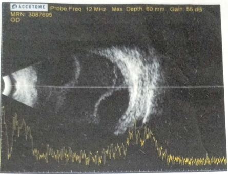
We present the case of a 29-year-old female who was recently diagnosed with diabetes mellitus. She presented to the eye department with sudden redness and increased watering (figure 1) in her left eye. On further investigation (B-Scan), she was diagnosed with endophthalmitis (figure 2) and managed with intravitreal antibiotics and topical antibiotics. During hospitalization, routine blood tests revealed a high random blood glucose level of 548 mg/dL and beta hydroxybutarate was low 0.01 mmol/L. However, she had no osmotic symptoms at that time. Intravenous regular insulin was administered, which resulted in a significant reduction in blood glucose levels.
Figure 1: Endophthalmitis
Figure 2: B-Scan of Left Eye.
Further investigation revealed that the patient had a history of childhood vision difficulties and had undergone surgery for lens dislocation, which was complicated by left eye glaucoma. She had poor school performance with no history of seizures, skin rash, or bony deformity. She had a family history of diabetes and was a product of consanguineous marriage.
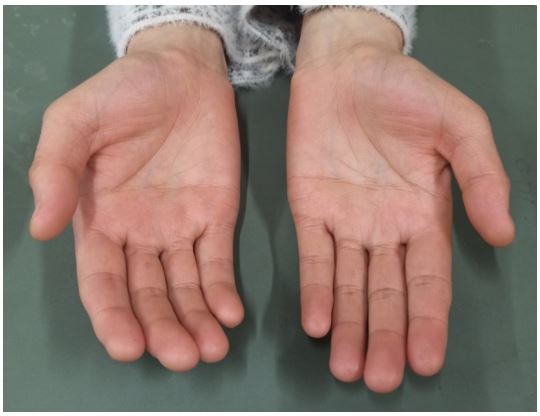
Figure 3: Arachnodactyly.
Figure 4: Scoliosis on X-ray.
Physical examination showed fare skin, high palate arch, thin long bone, arachnodactyly (figure 3), mental retardation (FSIQ was 60), osteoporosis (T-score was -3.0 on DEXA), hallux valgus, and scoliosis on x-ray (figure 4). Her ABG showed nonketotic metabolic acidosis, and plasma homocysteine levels were elevated (208.50 umol/L). 24 hour urine protein was nil, Vitamin D2 level was 10.9 ng/ml. Grade 1 pulmonary hypertension on ECHO. Color Doppler of both lower limb and Ultrasound abdomen was normal. Genetic testing could not be done due to financial constraints. The patient was managed with insulin on an MSI scale, pyridoxine 80 mg OD, folic acid 5 mg OD, Vitamin D3 60,000 IU weekly and intravitral and topical antibiotics under ophthalmologist consultation. With dietary modifications her blood glucose levels were closely monitored, and insulin dosage was adjusted accordingly.
Discussion
Homocysteinuria is a rare metabolic disorder caused by a deficiency of enzymes involved in the metabolism of methionine. It leads to the accumulation of homocysteine, which is toxic to blood vessels and other tissues. Homocysteinuria can present with a variety of symptoms, including intellectual disability, bone abnormalities, and vision problems. In this case, the patient had a history of childhood vision difficulties and underwent surgery for lens dislocation, which was complicated by left eye glaucoma. Elevated plasma homocystein levels have been associated with the development of diabetes and may have contributed to the patient's newly diagnosed diabetes [4]. Elevated plasma homocysteine levels, which may have contributed to the development of endophthalmitis as the initial manifestation of diabetes.
Conclusion
This case report highlights the importance of considering homocysteinuria in patients presenting with endophthalmitis in aphakic eyes and diabetes. It also underscores the need for close monitoring of blood glucose levels and insulin therapy in patients with homocysteinuria and diabetes. Further research is needed to determine the relationship between homocysteinuria, diabetes and endophthalmitis.
References
- Ueland PM, Refsum H. Plasma homocysteine, a risk factor for vascular disease: plasma levels in health, disease, and drug therapy. J Lab Clin Med. 1989; 114: 473.
- McCully KS. Homocysteine and vascular disease. Nat Med. 1996; 2: 386.
- Robinson K, Arheart K, Refsum H, et al. Low circulating folate and vitamin B6 concentrations: risk factors for stroke, peripheral vascular disease, and coronary artery disease. European COMAC Group. Circulation. 1998; 97: 437.
- Ala OA, Akintunde AA, Ikem RT, Kolawole BA, Ala OO, Adedeji TA. Association between insulin resistance and total plasma homocysteine levels in type 2 diabetes mellitus patients in south west Nigeria. Diabetes Metab Syndr. 2017; 11 Suppl 2: S803-S809. doi: 10.1016/j.dsx.2017.06.002. Epub 2017 Jun 10. PMID: 28610915.


