Clinical Image - Volume 3 - Issue 4
De novo formation of a giant dural arteriovenous fistula after cerebral venous sinus thrombosis
Sara Lima1; Ana Marques1; Andreia Matas1,2*
1Neurology Department, Centro Hospitalar de Trás-os-Montes E Alto Douro, Hospital de Vila Real, Vila Real, Portugal.
2Faculdade das Ciências da Saúde, Universidade da Beira Interior, Covilhã, Portugal.
Received Date : June 13, 2023
Accepted Date : July 10, 2023
Published Date: July 17, 2023
Copyright:© Andreia Matas 2023
*Corresponding Author : Andreia Matas, Neurology Department, Centro Hospitalar de Trás-os-Montes E Alto Douro, Hospital de Vila Real, Vila Real, Portugal.
Email: afmatas@chtmad.min-saude.pt
DOI: Doi.org/10.55920/2771-019X/1496
Clinical Image
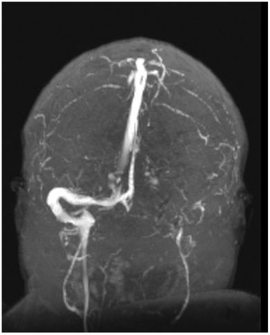
A 45-year-old male patient with past medical history of obesity and diabetes presented with complaints of pulsatile, frontal headache evolving in the past two days. His vital signs were normal and he had no meningeal signs or neurologic deficits. Magnetic resonance angiography (MRA) revealed cerebral venous sinus thrombosis (CVST) with intraluminal thrombus in galen's vein, and right rectum and transverse sinuses (Figure 1).
Figure 1: Magnetic resonance angiography (MRA) documenting cerebral venous thrombosis with intraluminal thrombus in galen's vein, and right rectum and transverse sinuses.
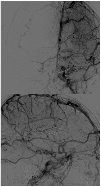
The patient was administered oral anticoagulation for six months and a brain MRA was repeated with recanalization of the previously documented venous thrombosis. Nevertheless, he evolved with daily headache, associated with nausea, dizziness and blurred vision. Fundoscopy revealed bilateral papilledema. A lumbar puncture showed raised intracranial pressure (opening pressure of 40 cm H20) without any abnormalities in the CSF analysis. The patient lost weight and initiated treatment with acetalozamide 1000 mg orally twice a day with no benefit. Lumbar puncture was repeated showing no reduction of CSF opening pressure. Brain MRA performed 18 months after the CVST revealed arterialization of the sigmoid sinus and part of the superior longitudinal sinus on the right and profusion of cortical veins and extracranial veins. Digital Subtraction Angiography (DSA) disclosed a dural arteriovenous fistula (DAVF) along the right lateral sinus and torcula and interesting the proximal aspect of the superior sagittal sinus, with anterograde venous drainage through the right sigmoid sinus, retrograde drainage to the left lateral sinus, superior sagittal sinus and straight sinus, and reflux into cortical veins (type IIa+b DAVF of Cognard classification) (Figure 2).
Figure 2: Digital Subtraction Angiography revealing a giant dural arteriovenous fistula along the right lateral sinus and torcula and also interesting the proximal aspect of the superior sagittal sinus, with anterograde venous drainage through the right sigmoid sinus, retrograde drainage to the left lateral sinus, superior sagittal sinus (including high convexity cortical veins) and straight sinus, and reflux into cortical veins (type IIa+b DAVF of Cognard classification).
DAVFs are unusual intracranial malformations that constitute up to 10-15% of cerebral malformations [1]. Although physiopathology underling DAVFs is largely unknown, it is possible that venous hypertension caused by impaired venous outflow in the context of head trauma, infections, tumors or CVST is implied [2,3]. This case highlights a rare cause of DAVF development following CVST.
References
- Gandhi D, Chen J, Pearl M, Huang J, Gemmete JJ, Kathuria S. Intracranial dural arteriovenous fistulas: Classification, imaging findings, and treatment. Am J Neuroradiol. 2012; 33(6): 1007-1013. doi:10.3174/ajnr.A2798
- Huang X, Shen H, Fan C, Chen J, Meng R. Clinical characteristics and outcome of dural arteriovenous fistulas secondary to cerebral venous sinus thrombosis: a primary or secondary event? BMC Neurol. 2023; 23(1): 131. doi:10.1186/s12883-023-03141-6
- Kusaka N, Sugiu K, Katsumata A, Nakashima H, Tamiya T, Ohmoto T. The importance of venous hypertension in the formation of dural arteriovenous fistulas: A case report of multiple fistulas remote from sinus thrombosis. Neuroradiology. 2001; 43(11): 980-984. doi:10.1007/s002340100596


