Case report - Volume 3 - Issue 4
Bronchopulmonary sequestration: A rare congenital anomaly presented in an adult female patient
Niaz Hussain; Hina Khalid*; Maham Zehra Zaidi; Kinza Zainab; Ghousia Aslam
Department of Surgery, Dow University Hospital Ojha campus DUHS, Karachi , Pakistan.
Received Date : June 13, 2023
Accepted Date : July 10, 2023
Published Date: July 17, 2023
Copyright:© Hina Khalid 2023
*Corresponding Author : Hina Khalid, Department of Surgery, Dow University Hospital Ojha campus DUHS, Karachi , Pakistan.
Email: hina.khalid@duhs.edu.pk
DOI: Doi.org/10.55920/2771-019X/1497
Abstract
Pulmonary sequestration is a congenital anomaly of lung parenchyma without a normal connection to the tracheobronchial tree and an anomalous systemic arterial supply. The presentation of this condition is variable ranging from no symptoms to hemoptysis. We report a case of pulmonary sequestration in an adult female who presented with recurrent chest infections. Her chest x-ray showed non-homogenous opacity in left lower lobe of lung and CT scan findings were suggestive of intralobar pulmonary sequestration in left lower lobe having arterial blood supply from the descending aorta and venous drainage in left inferior pulmonary vein. She subsequently underwent resection of the left, lower lobe of the lung.
Introduction
Bronchopulmonary sequestration represents a spectrum of abnormalities [1]. Pulmonary sequestration refers to the situation whereby a portion of lung tissue receives its blood supply from an anomalous systemic artery [2]. Two types of pulmonary sequestration are recognized [3-10], depending on whether or not the malformation possesses its own pleural covering. Intralobar sequestration is an abnormal region within the normal pulmonary parenchyma without its own pleural covering. Extralobar sequestration corresponds to a true accessory lung, with its own pleural envelope. Intralobar sequestration accounts for 0.15% to 1.7% of all congenital lung abnormalities [11]. Numerous reports have described serious complications arising from both intralobar as well as extralobar pulomonary sequestration such as fungal infections, tuberculosis, fatal hemoptysis, massive hemothorax, cardiovascular problems, benign tumors and even malignant degeneration [12]. We report this case to increase awareness about the condition and to highlight the significance of considering this rare etiology in a differential diagnosis of adult patients who present with recurrent pulmonary infections.
Case Report
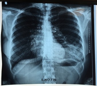
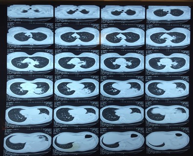
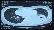
27 years old female presented in OPD with complaints of left sided chest pain, productive cough off and on for 2 years. She had a history of recurrent respiratory tract infections. She was previously diagnosed as having tuberculosis and had taken 2 courses of Antituberculous therapy, 13, and 11 years back, with no resolution of symptoms. On examination patient had mild anemia with dull percussion note and decreased air entry on left side of lower chest. The chest radiograph showed non homogenous opacity in the left lower zone of chest. CT scan showed multiple cystic spaces in left lower lobe with thin enhancing septae. Air specks were seen suggestive of infection. Enlarged paraesophageal, left hilar and perivascular lymph nodes were also seen. Findings were suggestive of intralobar pulmonary sequestration in left lower lobe having arterial blood supply from the descending aorta behind the left crus of diaphragm and venous drainage in left inferior pulmonary vein. No definite communication with tracheobronchial tree was seen. Overall appearance was suggestive of intralobar pulmonary sequestration with superimposed infection. A diagnosis of Left lower lobe type 3 pulmonary sequestration with bronchiectasis was made.
Figure 1: Chest x- ray showing non homogenous opacity in left lower zone.
Figure 2: CT-scan chest showing non homogenous opacity in left lobe of lung.
Figure 3: CT-scan showing multiple cystic spaces in left lower lobe.
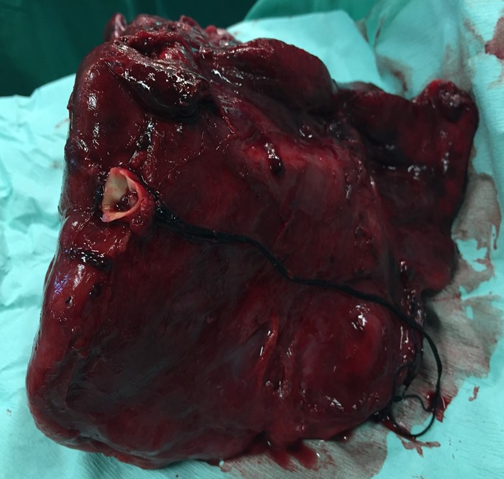
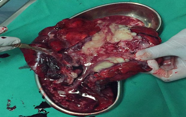
After pre-operative workup and anesthesia fitness, left thoracotomy and left lower lobectomy was performed. Per-operatively thick and discolored lower lobe of left lung with pus filled cystic spaces were found. The aberrant systemic artery of approximately 1cm caliber arising directly from aorta entering the posterior segment of lower lobe of the left lung was ligated. Post operatively patient remained stable, and the chest roentgenogram was within normal limits and patient was discharged on 10th postoperative day.
Figure 4: Showing resected left lower lobe of lung.
Figure 5: Showing sequestered tissue inside the left lower lobe of lung.
Discussion
Pulmonary sequestration is a relatively rare entity comprising 0.15%-0.4% of all congenital malformations.13 It occurs when a disturbance in embryonic development produces a cystic mass of non-functioning lung tissue. Most often the mass is supplied by an anomalous artery and has its own bronchial system, which usually does not communicate with the normal bronchial tree [14]. Pryce for the first time described this as a condition in which an abnormal artery is associated with an ectopic pulmonary mass in the lower lobe of lung [15].
Sequestrations are classified basically as, intralobar and extralobar. Intralobar according to the Pryce is classified into 3 types. Type 1 consists of regularly ventilated lung tissue perfused by two arterial blood supplies on the margins (pulmonary artery, systemic artery), type 2 of sequestered, irregularly ventilated (atelectatic) lung tissue perfused by two arterial blood supplies on the margins (pulmonary artery, systemic artery) and type 3 of sequestered, not ventilated lung tissue perfused only by the systemic artery blood supply.
Intralobar sequestration usually presents in adulthood and most commonly involves of left lower lobe [16,17]. Occasionally it is an incidental finding on roentgenograms of the chest in an asymptomatic patient; however the disorder is usually symptomatic and the most common presentation is recurrent pulmonary infections [18]. Plain chest radiograph is usually nonspecific, showing an ill-defined consolidation that mimics pneumonia, or shows a solitary soft tissue mass or nodule, or a cystic or multicystic lesion [19,20]. Chest CT usually shows a discrete mass in the posterior- or medial-basal segment of the lower lobe, with (as in our case) or without cystic changes [21]. Our patient also presented in a way intralobar pulmonary sequestration commonly presents. The standard treatment is resection of the segment or lobe that contains the sequestered tissue; the prognosis is favorable [22,23].
Conclusion
Bronchopulmonary sequestration can present with multiple nonspecific findings. This case illustrates a typical presentation of an intralobar bronchopulmonary sequestration and highlights the importance of considering this rare congenital condition when treating recurrent chest infection in young adults so that timely identification and management could be undertaken.
References
- Bhalla AS, Gupta P, Mukund A, Gupta M. Anomalous systemic artery to a normal lung: a rare cause of hemoptysis in adults. Oman Med J. 2012; 27(4): 319-22.
- Cornett HJ, Humphrey GM. Pulmonary sequestration. Paediatric respiratory reviews. 2004; 5(1): 59-68.
- Gdanietz K, Vorpahl K, Piehl G, Hock A. Clinical symptoms andtherapy of lung separation. Prog Pediatr Surg. 1987; 21: 86-97.
- Sade MR, Clouse M, Ellis H. The spectrum of pulmonarysequestration. Ann Thorac Surg. 1974; 18: 644-658.
- Boumghar M, Besson A, Saegeseer F. Sequestration pulmonaireintralobaire et surinfection tuberculeuse. Schweiz. Med. Wschr. 1979; 109: 1460-1464.
- Osada, H., Yokote, K., Arakawa, H., Yamate, N. Bilateral intralobarpulmonary sequestration. J Cardiovasc Surg. 1995; 1995: 36: 611-613.
- Saegesser F. Sequestrations pulmonaires. Schweiz Med Wschr. 1968; 49: 1919-1927.
- Leijala M, Louhimo I. Extralobar sequestration of the lung inchildren. Prog Pediatr Surg. 1987; 21: 98-106.
- Landing BH. Congenital malformations and genetic disorders of therespiratory tract. Am P Resp Dis. 1979; 120: 151-184.
- Rodgers MB, Harman PK, Johnson AM. Bronchopulmonary foregutmalformations. Ann Surg. 1986; 203: 517-524.
- Prasad R, Garg R, Verma SK. Intralobar sequestration of lung. Lung India. 2009; 26(4): 159.
- Van Raemdonck D, De Boeck K, Devlieger H, Demedts M, Moerman P, Coosemans W, Deneffe G, Lerut T. Pulmonary sequestration: a comparision between pediatric and adult patients. European Journal of Cardio-Thoracic Surgery. 2001; 19(4): 388-95.
- Loscertales J, Congregado M, Arroyo A, Jimenez-Merchan R, Giron JC, Arenas C, Ayarra J. Treatment of pulmonary sequestration by video-assisted thoracic surgery(VATS). Surgical endoscopy and other interventional techniques. 2003; 17(8): 1323.
- Carter R. Pulmonary sequestration. The annals of thoracic surgery. 1969; 7(1): 68-88.
- Pryce DM, Holmes Sellors T, Blair LG. Intralobar sequestration of lung associated with an abnormal pulmonary artery. British Journal of Surgery. 1947; 35(137): 18-29.
- Savic B, Birtel FJ, Tholen W et al. Lung sequestration: report of seven cases and review of 540 published cases. Thorax. 1979; 34(1): 96-101.
- Yadav A, Marthur RM, Devgarha S, Abraham VJ, Sisodia A, Goyal G. Intralobar pulmonary sequestration in right lower lobe with secondary infection in an adult male. Egyptian Journal of Chest Diseases and Tuberculosis. 2014; 63(1): 273-5.
- O’mara CS, Baker RR, Jeyasingham K. Pulmonary sequestration. Surgery, gynecology and obstetrics. 1978; 147(4): 609-16.
- Wei Y and Li F. Pulmonary sequestration: a retrospective analysis of 2625 cases in China. Eur J Cardiothorac Surg. 2011; 40: e39-42.
- Walker CM, Wu CC, Gilman MD, Godwin JD 2nd, Shepard JA and Abbott GF. The imaging spectrum of bronchopulmonary sequestration. Curr Probl Diagn Radiol. 2014; 43: 100-114.
- Kang M, Khandelwal N, Ojili V, Rao KL, Rana SS. Multidetector CT angiography in pulmonary sequestration. Journal of computer assisted tomography. 2006; 30(6): 926-32.
- Frazier AA, Rosado de Christenson ML, Stocker JT, Templeton PA. Intralobar sequestration: radiologic-pathologic correlation. Radiographics. 1997; 17(3): 725-745.
- Hirai S, Hamanaka Y, Mitsui N, Uegami S, Matsuura Y. Surgical treatment of infected intralobar pulmonary sequestration: a collective review of patients older than 50 years reported in the literature. Ann Thorac Cardiovasc Surg. 2007; 13(5): 331-334.

