Case report - Volume 3 - Issue 4
Bithalamic signal anomaly
El Houss Salma*; Laasri Khadija; Amsiguine Najwa; El Fenni Jamal; Edderai Meryem
Radiology Department, Mohammed V Military Training Hospital, Rabat, Morocco.
Received Date : June 16, 2023
Accepted Date : July 14, 2023
Published Date: July 21, 2023
Copyright:© El Houss Salma 2023
*Corresponding Author : El Houss Salma, Radiology Department, Mohammed V Military Training Hospital, Rabat, Morocco. Tel: +212 642294009.
Email: salma.elhous94@gmail.com
DOI: Doi.org/10.55920/2771-019X/1504
History
40 year old patient with acute headaches for 3 days, complicated by an epileptic seizure disorder.
Diagnosis
Thrombophlebitis of the internal cerebral veins
Commentary
Deep veinous thrombosis is manifested by headache, vomiting, neurological deficit or confusional state. In more severe forms and due to the function of the affected venous territory, it manifests itself by consciousness disorders. The diagnosis is based on imaging. In cases in which a CT scan has been performed as a first-line procedure, it shows both direct and indirect signs [1].
Direct signs include spontaneous hyperdensity of the thalamic veins in contact with the interventricular foramen, basal veins of Rosenthal at the level of an ambient cistern, internal cerebral veins, vein of Galen, or the right sinus. After intravenous injection of iodinated contrast, thrombosis is recognized as a defect in a deep vein. Indirect signs are hypodensity of the thalamus that may extend to the gray nuclei of the diencephalon, the deep white matter and the posterior part of the midbrain [2].
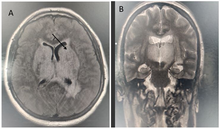
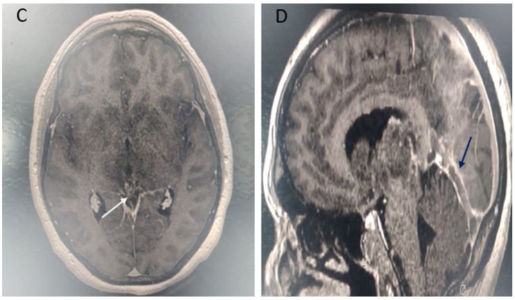
Conventional MRI and MR angiography are more efficient than CT. The thrombus is easier to visualize because it is hyperintense in T1 and T2 After intravenous injection of gadolinium, it appears in hyposignal surrounded by enhancement of the walls of the Dural (C , white arrow and D, blue arrow), and hyperintensity of the thalami that may extend to the gray nuclei of the diencephalon (A and B)
Figure 1: T2 weighted sequence demonstrate an hyperintensity of the thalami that may extend to the gray nuclei of the diencephalon (A and B)
Figure 2: T1 weighted sequence with PDC injection demonstrate an hyposignal surrounded by enhancement of the walls of the Dural (C , white arrow and D, blue arrow), in relation to The thrombus
References
- Gaillard F, Guan H, Ashraf A, et al. Cerebral venous thrombosis. Reference article, Radiopaedia.org (Accessed on 02 May 2023) https://doi.org/10.53347/rID-4449.
- Bonneville, Imagerie des thromboses veineuses cérébrales. 2014; 4333(12): 1113-1181. ISSN 2211-5706. http://dx.doi.org/10.1016/j.jradio.2014.09.005,(http://www.sciencedirect.com/science/article/pii/S2211-5706(14)00421-4


