Review Artcile - Volume 3 - Issue 4
Percutaneous Internal Ring Suturing (Pirs): A minimally invasive procedure in pediatric inguinal hernia repair
Mohamed Shafi Bin Mahboob Ali*
Advanced Medical and Dental Institute, Malaysia.
Received Date : June 21, 2023
Accepted Date : July 19, 2023
Published Date: July 26, 2023
Copyright:© Mohamed Shafi Bin Mahboob Ali 2023
*Corresponding Author : Mohamed Shafi Bin Mahboob Ali, Advanced Medical and Dental Institute, Malaysia.
Email: mshafix_7@yahoo.co.uk
DOI: Doi.org/10.55920/2771-019X/1509
Abstract
Purpose: Percutaneous internal ring suturing (PIRS) is a single-port laparoscopic technique to repair a pediatric indirect inguinal hernia. We used PIRS in our babies and found that the surgery time was shortened, with smaller incisions, lesser complications, and early discharges. This is a retrospective case study to introduce our novel minimally invasive method of repairing pediatric hernias with fewer complications, safe and cost-effective.
Methods: Healthy-term male babies with indirect inguinal hernia and a thin abdominal wall were chosen. After general anesthesia, a one-cm infra-umbilical incision was made, and a 3mm 70◦ scope was inserted after a Pneumoperitoneum. With the guidance of the scope, a spinal needle with a Prolene 2-0 was punctured and threaded in on the medial edge of the internal inguinal ring. Once the suture is in, the needle is withdrawn. Another Prolene 2-0 is punctured and threaded on the lateral edge of the internal inguinal ring. The needle is looped into the first suture. The sutures were tightened and ligated. A laparoscope is used to inspect that the internal ring is snugly closed without the entrapment of the cord structures. The scope is removed, and the port site is sutured. The baby was extubated and observed in the general ward.
Results: PIRS is a simple pediatric inguinal hernia repair technique with shortened operative time, lesser scar and pain, early recovery, and discharge.
Conclusion: The PIRS technique is safe, with lesser complications and satisfactory results.
Keywords: Percutaneous, internal, suturing, hernia
Introduction
A congenital inguinal hernia in the pediatric population is one of the most common conditions requiring surgical treatment, with the reported incidence ranging between 1% and 5% [1, 2, and 3]. It manifests clinically more commonly in younger children, especially premature infants, and the incidence rate is up to 10 times higher in boys. Percutaneous internal ring suturing (PIRS) is a single-port laparoscopic technique used to repair a pediatric indirect inguinal hernia [4]. This technique is still new in the fraternity, and only a few centers practice it. We applied this technique to our patients and found that the duration of surgery was shortened, with smaller surgical incisions, lesser complications and early discharge. Laparoscopic single port needle-assisted hernia repair with percutaneous internal ring suturing (PIRS) technique is frequently preferred to repair inguinal and diaphragmatic hernias [5, 6]. This case aims to present laparoscopic single port needle-assisted repair of pediatric hernias done in our center.
Methodology
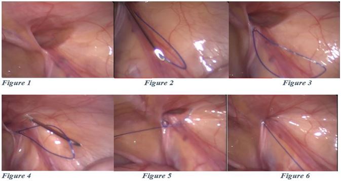
Patient selection was made based on several criteria, term baby with no anomalies, indirect inguinal hernia, and male baby with a thin abdominal wall. The procedure was performed under general anesthesia. Pneumoperitoneum was established using an open technique through the umbilicus, and a 5-mm trocar was placed [Figure 1]. A 1-mm incision was then made with an 11-size scalpel just above the inner opening of the inguinal canal. Special care was taken to avoid injury to the spermatic cord and testicular vessels while catching the peritoneum from a 12 o’clock position to an 8 o’clock position in correct sided operations, leaving the loop in situ[Figure 2]. Once the sutures are in, the needle is withdrawn [Figure 3]. Another Prolene 2-0 is punctured and threaded in on the lateral edge of the internal inguinal ring [Figure 4]. This time, the needle is looped into the first suture [Figure 5]. After the withdrawal of the needle, the sutures were tightened and ligated [Figure 6]. Laparoscope used to inspect that the internal ring is snugly closed without the entrapment of the vas or the cord structures. Once ligated, the scope is removed, and the port site is sutured using a purse string suture. Fascial closure at the umbilicus was done using polyglactin thread. Skin closure was performed using either histoacryl adhesive or absorbable intracuticular suture.
Results
Demographic data and clinical evaluation findings
Of the 80 patients, 50 (62.5%) were female, and 30 (37.5%) were male, with higher number of patients (60 (75%)), coming from the rural area. The median age of the patients was 106 (range, 5–204) months. Twenty-nine patients (36.3%) were aged between 0 - 48 months, 20 (25%) were aged between 60-108 months, and 31 (38.8%) were aged between 120-216 months. On clinical findings and laboratory examinations, the most common symptom at presentation was the presence of neck swelling, which was found in 30 (37.5%) patients. Fever and night sweating were present in 10 patients (8%), restricted movement and pain in the extremities were present in 9 (7.2%), the cough was present in 11 (8.8%), hematuria was present in 10 (8%), and weight loss was present in 5 (4%) patients. Blurred consciousness, vomiting, and headache were found in 2 (1.6%), vomiting was found in 1 (0.8%) patients, and growth and developmental delay was found in 2 patients (1.6%). As a result of history and family screening, contact with an individual with tuberculosis in the immediate vicinity was found in 27 (33.7%) patients, as shown in Table 1
PIRS technique:
Figure 1: Right inguinal hernia— internal inguinal ring.
Figure2: The same needle was introduced at same skin incision but on the other side of the internal ring.
Figure 3: Introduction of a non-absorbable monofilament nylon loop through half of the inguinal ring.
Figure 4: The needle was passed through the previously introduced loop and suture is pushed through the needle.
Figure 5: After removal of the needle, the loop was drawn out and the knot passed around internal ring.
Figure 6: Closed internal ring.
Results
PIRS is a straightforward technique in pediatric inguinal hernia repair with shortened operative time, lesser scar and postoperative pain with early recovery. We performed only 2 cases of recurrence in the PIRS group of patients. No cases of testicular atrophy were reported in all our cases. There was only 1 case of surgical site infection at the wound site, where we started the baby on antibiotics and were subsequently discharged home. Otherwise, no cases of hernia recurrence, hydrocele or hematoma were reported during the regular clinic follow-up.
Discussion
In our study, the PIRS technique had shorter operative and theatre times and fewer complications than the traditional open surgical method. It has been understood that the laparoscopic approach to inguinal hernia repair is more expensive, consumes more instruments and is expensive compared to conventional open surgery. Our studies found that the PIRS method took less time with fast recovery and early discharge compared to the open herniorrhaphy. No perioperative complications occurred, and the number of postoperative complications was less for the PIRS group. PIRS also has the potential to save operating time, especially for patients with bilateral hernias. Peng has made a fascinating observation, similar to ours, that the duration of left-sided hernia surgeries is longer than right-sided ones using the laparoscopic method [7]. It might be explained by the fact that left inguinal repair is more difficult for a right-handed person standing on the patient's right side. All surgeons were right-handed in our material. PIRS is claimed to be a safe treatment method in inguinal hernia repair and is preferred frequently [7, 8]. The advantage of this technique is the use of only one optical port and the lack of manipulation and suturing with instruments inside the peritoneal cavity. Placement of internal sutures on the internal inguinal canal opening can lead to natural tapering of the inguinal canal and no recurrence of the hernia, as seen in the umbilical hernia in neonates and infants. It was estimated that the incidence of a contralateral metachronous hernia was 9.62%, and the ipsilateral recurrent hernia was 1.23% [9, 10]. The incidence of a metachronous contralateral pediatric inguinal hernia is 6.4% in both genders. [11, 12, 13]
Conclusion
The percutaneous internal ring (PIRS) technique is straightforward and safe, with lesser complications and satisfactory results. PIRS technique allows for a quick return to full fitness of children. It does not leave visible scars, and the complication rate is not significantly higher than after conventional surgery.
References
- Ravi K. Hamer, D.B. Surgical treatment of inguinal herniae in children. Hernia. 2003; 7: 137-140.
- Rosenberg, J. Pediatric inguinal hernia repair—A critical appraisal. Hernia 2008; 12: 113-115.
- Esposito C, St Peter SD, Escolino M, Juang D, Settimi A, Holcomb GW. 3rd. Laparoscopic Versus Open Inguinal Hernia Repair in Pediatric Patients: A Systematic Review. J. Laparoendosc Adv. Surg. Tech. 2014; 24: 811-818.
- Shalaby R, et al. One trocar telescopic assisted inguinal hernia repair in children: a novel technique. J Pediatr Surg. 2017; 53(1):192-8.
- Yilmaz E, et al. A novel technique for laparoscopic inguinal hernia repair in children: single-port laparoscopic percutaneous extraperitoneal closure assisted by optical forceps. Pediatr Surg Int. 2015; 31(7):639-46
- Peng Y, Li C, Han Z, Nie X, Lin W. Modified single-port vs two-port laparoscopic herniorrhaphy for children with concealed deferent duct: A retrospective study from a single institution. Hernia 2017; 21: 435-441
- Yilmaz E, et al. A novel technique for laparoscopic inguinal hernia repair in children: single-port laparoscopic percutaneous extraperitoneal closure assisted by optical forceps. Pediatr Surg Int. 2015; 31(7): 639-46
- Stone ML, et al. Novel laparoscopic hernia of Morgagni repair technique. J Thorac Cardiovasc Surg. 2012; 143(3): 744-5
- Chin TW, Pan ML, Lee HC, Tsai HL, Liu CS. Second hernia repairs in children—a nationwide study. J Pediatr Surg. 2015; 50: 2056-2059. doi: 10.1016/j.jpedsurg.2015.08.024
- Nataraja R, Mahomed A. Metachronous contralateral pediatric inguinal hernia. Open Access Surgery. 2010; 3: 87-90
- Watanabe T, Yoshida F, Ohno M, et al. Morphology-based investigation of metachronous inguinal hernia after negative laparoscopic evaluation - is it acquired indirect inguinal hernia? J Pediatr Surg. 2016; 51: 1548-1551. doi: 10.1016/j.jpedsurg.2016.03.008
- Miyake H, Fukumoto K, Yamoto M, et al. Risk factors for recurrence and contralateral inguinal hernia after laparoscopic percutaneous extraperitoneal closure for pediatric inguinal hernia. J Pediatr Surg. 2017; 52(2): 317-321. doi: 10.1016/j.jpedsurg.2016.11.029
- Lee SR, Choi SB. The efficacy of laparoscopic intracorporeal linear suture technique as a strategy for reducing recurrences in pediatric inguinal hernia. Hernia. 2017; 21: 425-433. doi: 10.1007/s10029-016-1546-y


