Clinical Image - Volume 3 - Issue 4
Brownish limb tumor as a presentation of primary hyperparathyroidism
Patricia Agüero1; Nadia Arguiñarena2; Gabriela Mintegui3*
1Adjunct Professor, Clinic of Endocrinology and Metabolism. School of Medicine, Hospital of Clinics, Uruguay.
2Resident, Clinic of Endocrinology and Metabolism, School of Medicine. Hospital of Clinics, Uruguay.
3Associate Professor, Clinic of Endocrinology and Metabolism, School of Medicine, Hospital of Clinics, Uruguay.
Received Date : June 28, 2023
Accepted Date : July 21, 2023
Published Date: July 28, 2023
Copyright:© Gabriela Mintegui 2023
*Corresponding Author : Gabriela Mintegui, Clinic of Endocrinology and Metabolism, Associate professor, School of Medicine, Hospital of Clinics, Uruguay.
Email: gabymin92@gmail.com
DOI: Doi.org/10.55920/2771-019X/1514
Clinical Image
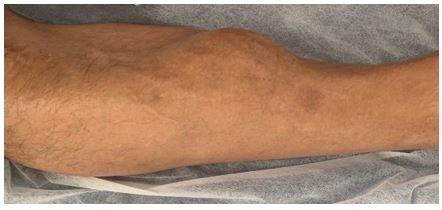
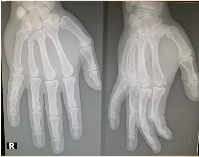
43-year-old male, with tumor in the anterior region of the right lower limb (figure 1) and biopsy compatible with brown tumor. He presented total serum calcium 12.3 mg/dl and 13 mg/dl (NR: 8.3-10.3), parathormone (PTH) 1057 pg/ml (NR 15-65). With the diagnosis of primary hyperparathyroidism, a 99mTc-MIBI parathyroid scintigraphy with SPECT was requested, which showed hyperfunctioning parathyroid tissue in the projection of the left inferior parathyroid gland. Hand radiographs showed a lytic lesion in punch in the distal phalanx of the first finger of the left hand (figure 2); in the clavicle radiograph: lytic lesion of 10 mm x 8 mm in the right clavicle, no alterations were evidenced in the skull radiograph.
Symptomatic primary hyperparathyroidism is a rare presentation today. In severe metabolic osteopathy (osteitis fibrosa cystica), the following may be observed: bone cysts, brown tumors, and fibrosis of the marrow spaces. Bone involvement is predominantly cortical bone, with greater deterioration at the level of the distal third of the radius; although the trabecular bone may be compromised, with a greater risk of fractures in those sites [1]. Cystic osteitis fibrosa cystica is due to the action of PTH in the bone and is currently rare as an initial presentation of primary hyperparathyroidism. Given the access to serum calcium determination, it is diagnosed in early stages of this disease asymptomatically. Clinically it may present with bone pain, bone deformities and pathological fractures, much more rarely with a visible tumor as in this case. With treatment of primary hyperparathyroidism, brown tumors may disappear [2].
In conclusion, brown tumor is a rare form of initial presentation of hyperparathyroidism at present. Treatment is resection of the parathyroid adenoma as it usually regresses with decreasing PTH.
Figure 1: Tumor in the anterior region of the right lower limb.
Figure 2: Hand radiographs showed a lytic lesion in punch in the distal phalanx of the first finger of the left hand.
References
- Mintegui G, Mendoza B. Hiperparatiroidismo primario. Perspectiva actual. 2017; 51: 177-182.
- Mintegui G, Mendoza B. Brown tumor regression in parathyroid adenoma. 2020; 155(11): 519. https://doi.org/10.1016/j.medcli.2019.09.008.


