Case Report - Volume 3 - Issue 5
Clear cell sugar tumour – A rare lung tumour
João Paulo Duarte dos Santos Oliveira Rodrigues*, Mário Pinto, Miguel Cristóvão, Rita Gerardo
Centro Hospitalar e Universitário Lisboa Central, Portugal.
Received Date : July 26, 2023
Accepted Date : Sep 07, 2023
Published Date: Sep 14, 2023
Copyright: © Joã Paulo Duarte dosSantos Oliveira Rodrigues 2023
*Corresponding Author: João Paulo Duarte dos Santos Oliveira Rodrigues, Hospital de Santa Marta - Centro Hospitalar e Universitário
Lisboa Central, Portugal.
Email: joao.oliveira.rodrigues.pneumo@gmail.com"
DOI: Doi.org/10.55920/2771-019X/1547
Clinical Image
A 82-year-old woman, never smoker, retired farmer with known medical history of hypertension, heart failure, chronic kidney disease, hyperuricemia, and dyslipidaemia was referred to a pulmonology consultation due to the identification of a nodule on a chest-x-ray and one-year history of fatigue. A chest CT scan confirmed a well-defined nodular lesion (22x20mm) in the external segmental of the middle lobe. A PET-CT scan showed increased metabolic activity (SUVMax of 3.5). A transthoracic biopsy resulted in the histological identified epithelioid cells with a clear cytoplasm, and immunohistochemical positivity for HMB45 and Melan A confirming a Clear Cell Sugar Tumor or PEComa. In multidisciplinary discussion of thoracic oncology it was decided to proceed with imaging surveillance due to the presence of significant comorbidities and the typically indolent behaviour of the tumour. Patient underwent semi-annual imaging reevaulations that did not show any tomographic changes.
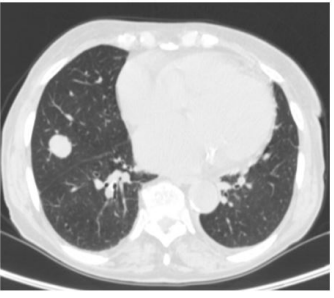
Figure 1: Septembter 2020- well defined nodule in the middle lobe.
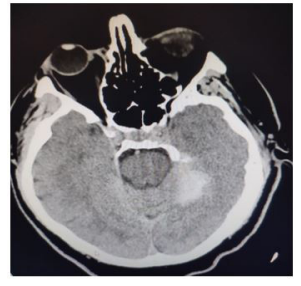
Figure 1: Brain CT of the patient at the admission showing a left sided hyperdense lesion on axial plane.
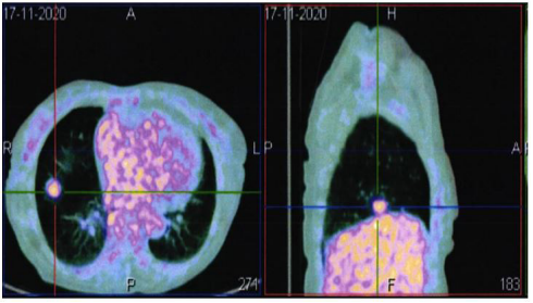
Figure 2: PET scan identifying a SUVMax 3,5
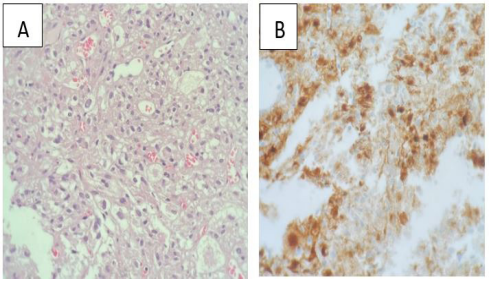
Figure 3: Panel A: Clear cells under hematoxylin and eosin staining.
Panel B: positivity for HMB 45.
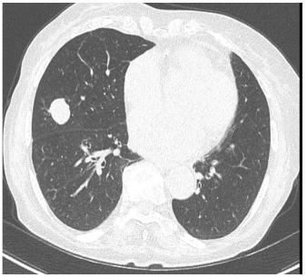
Figure 4: December 2021-stable disease.
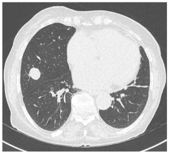
Figure 5: August 2022- stable disease.

