Case Report - Volume 3 - Issue 5
Bartholin’s gland cyst
Mejjati Kaoutar*; Soughi Meryem; Douhi Zakia; Elloudi Sara; Baybay Hanane; Mernissi Fatima Zahra
Department of Dermatology and Venereology, University Hospital Hassan II, Fez, Morocco.
Received Date : August 17, 2023
Accepted Date : Sep 18, 2023
Published Date: Sep 25, 2023
Copyright: © Mejjati Kaoutar 2023
*Corresponding Author: Mejjati Kaoutar, Department of Dermatology and Venereology, University Hospital Hassan II, Fez, Morocco.
Email: kaoutarme@hotmail.fr
DOI: Doi.org/10.55920/2771-019X/1554
Clinical Image
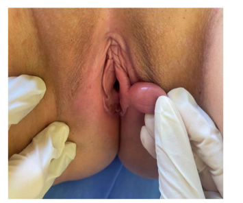
A 39-year-old woman with no notable pathological history presented to our department with a genital formation that had been progressively evolving for 1 year and becoming uncomfortable. The clinical examination found a painless nodule of cystic consistency measuring approximately 2 cm, covered with normal skin located in the labia minora (Figure 1). We performed an excisional biopsy under local anesthesia, revealing cystic formation with a regular, simplified cubo-cylindrical epithelium, resting on a fibrous layer. The lumen contained a polymorphous inflammatory infiltrate with cellular debris. The diagnosis of Bartholin’s cyst was made.
Bartholin's gland cysts are the most common vulvar cysts. They affect women particularly between the ages of 20 and 30. They result from ductal obstruction; the cause is usually unknown. Bartholin's cysts are generally asymptomatic, but can become infected and painful and form an abscess, which is known as bartholinitis. Diagnosis is clinical. They are painless to palpate, unilateral and palpable near the vaginal orifice. Treatment is based on excision in case of discomfort or recurrence.

Figure 1: Roundish nodule of cystic consistency covered with normal skin located in the labia minora.

