Case Report - Volume 3 - Issue 5
Hibernoma: An unveiling of a rare adipocytic tumor
Raghav Kapoor*, Prerna Chadha
Department of Pathology, Rajiv Gandhi Cancer Institute and Research Centre, New Delhi, Delhi, India.
Received Date : August 22, 2023
Accepted Date : Sep 22, 2023
Published Date: Sep 29, 2023
Copyright: © Raghav Kapoor 2023
*Corresponding Author : Raghav Kapoor, Department of Pathology, Rajiv Gandhi Cancer Institute and Research Centre, New Delhi, Delhi, India.
Email: kapoor.raghav1212@gmail.com
DOI: Doi.org/10.55920/2771-019X/1558
Introduction
Classic lipomas are among the most prevalent soft tissue tumors and histologically resemble white fat. The back, specifically the inter-scapular region, is the location of hibernoma that is most frequently observed. The neck, axillae, thigh, and intra-thoracic region are other frequent locations. Hibernoma is a lesion that shouldn't be excluded from the differential diagnosis because it can't be distinguished from malignant tumors clinically or radiographically.
Case Report
A 53-year-old female patient with a history large swelling at upper back region for 11 months came with the complaints of dull aching pain started 1 month back. There was no history of trauma and patient had overall good built and stature. On examination, a large and painful subcutaneous/intramuscular swelling at inter-scapular region was noted approximately ~9cm in largest dimension. The mass was mobile, not associated with any skin changes and erythema, non-pulsatile and non-tender. Imaging showed a 8.33 x 7.45 cm mass in intramuscular plane with well-defined borders and non-infiltrative nature. No evidence of necrosis was seen. An opinion consistent with Lipoma was given on radiology. However, HPE was advised to rule out Lipsosarcoma. On gross, it was capsulated, lobular yellow mass having a homogenous fatty cut surface. Areas of hemorrhage and necrosis were not seen.
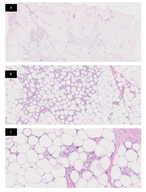
On microscopic examination, low power (A) shows a large adipocytic neoplasm. On medium power (B) tumor cells were seen comprising predominately large, pale, and eosinophilic brown fat cells with a tiny, multivacuolated central nucleus, interspersed with varying numbers of univacuolated white cells. Higher power (C) image shows no evidence of Cytological atypia, necrosis and mitosis was seen. A diagnosis of Benign Brown adipose tissue tumor, consistent with Hibernoma was given.
Discussion
Hibernoma Comprises < 2% of benign lipomatous tumors and makes up 1% of all adipocytic tumors. The thigh (29%), shoulder (12%), back (10%), neck (9%) and chest wall (6%), are the most frequent sites [2,3]. Other sites: intraabdominal, retroperitoneal and thoracic cavity [4]. Few cases show an association with MEN type 1 [5]. Imaging reveals a well-defined, hyperdense lesion that is occasionally intramuscular and subcutaneous which is indistinguishable from Liposarcoma [6].
Macroscopically, hibernomas are mobile, well defined, soft and on lipid concentration variability, color ranges from yellow to light brown to gray, with thin capsules. Microscopically, the tumor is characterized by brown fat having large multi-vacuolated cells with abundant mature adipocytes, several branching capillaries, and no significant atypia/mitosis/necrosis [1,7]. On immunohistochemistry, diffuse S100 positivity seen with very low Ki67 index [8].

References
- Furlong MA, Fanburg-Smith JC, Miettinem M. The morphologic spectrum of hibernoma: a clinicopathologic study of 170 cases. Am J Surg Pathol. 2001; 25(6): 809-814.
- Al Hmada Y, Schaefer IM, Fletcher CDM. Hibernoma Mimicking Atypical Lipomatous Tumor: 64 Cases of a Morphologically Distinct Subset. Am J Surg Pathol. 2018; 42(7): 951-957. doi: 10.1097/PAS.0000000000001061. PMID: 29629919; PMCID: PMC6438389.
- Shackelford RE, Al Shaarani M, Ansari J, Wei E, Cotelingam J. A Twenty-Four-Year-Old Woman with Left Flank Lipoma-Like Hibernoma. Case Rep Oncol. 2017; 10(2): 438-441. doi: 10.1159/000475708. PMID: 28626403; PMCID: PMC5471793.
- Tsuda Y, Matsuyama A, Makihara K, Higaki K, Motoi T, Okuma T, Hisaoka M. Nuclear expression of MDM2 in hibernoma: a potential diagnostic pitfall. Virchows Arch. 2021; 478(3): 527-534. doi: 10.1007/s00428-020-02914-5. Epub 2020 Aug 20. PMID: 32815035.
- Nord KH, Magnusson L, Isaksson M, Nilsson J, Lilljebjörn H, Domanski HA, Kindblom LG, Mandahl N, Mertens F. Concomitant deletions of tumor suppressor genes MEN1 and AIP are essential for the pathogenesis of the brown fat tumor hibernoma. Proc Natl Acad Sci U S A. 2010; 107(49): 21122-7. doi: 10.1073/pnas.1013512107. Epub 2010 Nov 15. PMID: 21078971; PMCID: PMC3000312.
- AlQattan AS, Al Abdrabalnabi AA, Al Duhileb MA, Ewies T, Mashhour M, Abbas A. A Diagnostic Dilemma of a Subcutaneous Hibernoma: Case Report. Am J Case Rep. 2020; 21: e921447. doi: 10.12659/AJCR.921447. PMID: 32332693; PMCID: PMC7200092.
- Lele Subodh M, Chundru Satish, Chaljub Gregory, Adegboyega Patrick, Haque Abida K. Hibernoma. Archives of Pathology & Laboratory Medicine. 2002; 126(8): 975-978.
- Song B, Ryu HJ, Lee C, Moon KC. Intraosseous Hibernoma: A Rare and Unique Intraosseous Lesion. J Pathol Transl Med. 2017; 51(5): 499-504. doi: 10.4132/jptm.2017.07.28. Epub 2017 Aug 22. PMID: 28827513; PMCID: PMC5611535.

