Case Report - Volume 3 - Issue 5
Carotid body tumor: A case report
Mribat M*; Bourekba I; El Adioui G; Lahkim M; En-Nouali H
Radiology Department, Mohammed V hospital, Mohammed V university Rabat, Morocco
Received Date : Aug 28, 2023
Accepted Date : Sep 29, 2023
Published Date: Oct 10, 2023
Copyright: © Mribat Merieme 2023
*Corresponding Author : Mribat Merieme, Radiology Department, Mohammed V hospital, Mohammed V university Rabat, Morocco.
Email: meriememribat@gmail.com
DOI: Doi.org/10.55920/2771-019X/1564
Abstract
Carotid body tumors (CBTs), also referred to as paragangliomas or chemodectomas, are rare neuroendocrine neoplasms arising from glomus cells near the carotid bifurcation. These tumors, constituting less than 1% of head and neck cancers, are often asymptomatic and detected incidentally through radiological imaging or clinical examination. These tumors pose diagnostic and management challenges due to their intricate location and potential vascular involvement. The preferred course of treatment is surgical excision. Preoperative embolization and surgical planning both benefit from preoperative angiography.
Introduction
Carotid body tumors, arising from the neural crest-derived paraganglia located at the carotid bifurcation, they’re rare representing less than 0,7% of head and neck neoplasms. Radiological techniques play a pivotal role in enhancing our understanding of the anatomical relationship between these tumors and adjacent structures, guiding treatment decisions, and improving patient outcomes. We present the case of a 60-year-old in whom CBT was diagnosed following hospitalization for a progressive cervical mass.
Case report
A 60-year-old individual with a cervical mass presented complaints of swelling on the right side of the neck persisting for 6 months, along with intermittent pain. There was no history of fever, cough, weight loss, or loss of appetite. The patient did not report any swelling in the bilateral axillary region, ptosis, hoarseness of voice, or syncopal attacks. Notably, the patient had no prior medical history of tuberculosis, or diabetes. Upon local examination, a distinct swelling was observed on the anterolateral aspect of the neck muscle, positioned just below the angle of the mandible on the right side of the neck . The swelling exhibited a firm-hard texture, slight tenderness, a minor pulsatile sensation, and slight mobility from side to side. There was no indication of localized temperature elevation. Laboratory tests, including complete blood count (CBC), thyroid function test (TFT), kidney function test (KFT), and liver function test (LFT), were all within normal ranges.
The patient was advised to undergo contrast-enhanced computed tomography (CECT) of the neck. The plain computed

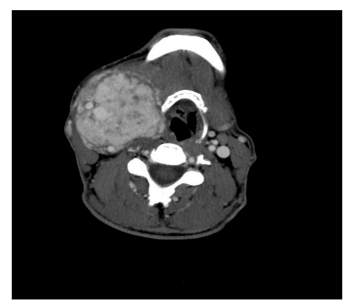
Figure 1: Axial cross-sectional computed tomography (CT) scan reveals a right lateral cervical multilobulated mass prominently encasing the carotid axis, exhibiting intense enhancement. No calcifications are seen.
tomography (CT) sagittal section revealed an indistinct mass lesion of soft tissue density on the right side of the neck, anterior to the SCM muscle, and inferior to the angle of the mandible. The mass extended superiorly up to the suprahyoid space . No calcifications were evident within the mass . The post-contrast study axial section demonstrated a multilobed mass lesion within the carotid sheath on the right side of the neck, at the bifurcation of the right CCA, leading to splaying of the ICA and ECA (Lyre's sign). The lesion exhibited intense and uniform enhancement.

Figure 2: Sagittal and coronal sections enable an improved visualization of the mass's relationship with the carotid axis. An enlargement of the bifurcation is discernible also called “Lyre’s sing”.
Discussion
A paraganglioma is a tumor that can develop anywhere in the body from paraganglia, which are non-neuronal cells descended from neural crest cells [5]. The carotid body, the vagus nerve in the head and neck region, the jugular foramen, the middle ear, the aortopulmonary window, and the organ of Zuckerkandl are the common sites. The manifestations of carotid body paraganglioma might be sporadic or familial. A lateral neck lump with modest growth and no pain is how this lesions frequently appear. Cranial nerve palsies, voice changes, and hearing loss maybe be the accompanying symptoms.
The majority of the time, CBTs are discovered accidentally on imaging studies or through clinical assessment. Image analysis and pathological anatomy play a major role in the final diagnosis. Doppler ultrasound, CECT neck, contrast enhance magnetic resonance imaging (MRI) neck, and angiography plays an important role in the diagnosis of CBT. The best screening method for CBTs is a color-flow carotid duplex, and CBT is characterized by a well-defined hypoechoic mass causing splaying of the ICA and ECA. On color Doppler imaging hypervascularity is seen within the lesion with a low-resistance flow pattern. CECT and contrast-enhanced MRI contribute to accurate diagnosis and surgical planning. CBTs exhibit intense enhancement and well-defined margins. On CT, they present as soft tissue density masses, while on MRI, they appear hypointense on T1-weighted images and hyperintense on T2-weighted images. Diffusion-weighted imaging (DWI) aids in differentiation, showing restricted diffusion with low apparent diffusion coefficient (ADC) values, in both CT and MRI the lesion show an intense enhancement. On digital subtraction angiography (DSA), a tumor with an "early vein" visible due to arteriovenous shunting is detected by a high blush and the splaying of the carotid vessels.
Surgical tumor resection depends on the relationship of the tumor with the artery bifurcation and the proximity of cranial nerves diagnosed accurately on CT angiography (CTA) or MR angiography (MRA). CTA can characterize the tumor's size and relationship to bony landmarks, which can change the surgical strategy. On dynamic contrast-enhanced MRI has better results for assessing the carotid and nerve involvement.

