Case Report - Volume 3 - Issue 5
A case report of a symptomatic pericardial cyst: Comprehensive review of diagnosis and management
Phan Thanh Ha Le1; Hoang Anh Tien1,2; Viet Tang Thai3; Ngoc Long Phi Ta4; Minh Hung Ngo3; Anh Binh Ho5;Nguyen Tri Nhan Le3; Van Phuoc Ho6; Nelson Pham7; Van Tuong Nguyen8; Phillip Tran3,9*
1Hue University of Medicine and Pharmacy, Hue Province, Vietnam.
2Internal Medicine Department, Hue University Vice Director of Cardiovascular Centre, Hospital of Hue University of Medicine and Pharmacy – Hue Province, Vietnam.
3Nam Can Tho University, Can Tho City, Vietnam.
4Tam Duc Hospital, Ho Chi Minh City, Vietnam.
5Cardiovascular Centre, Hue Central Hospital, Vietnam.
6Head of Interventional Cardiology Department, Da Nang Hospital, Da Nang City, Vietnam.
7University of Arizona College of Medicine, AZ, Vietnam.
8Pham Ngoc Thach University, Ho Chi Minh City, Vietnam.
9Cardiovascular Department, Yavapai Regional Medical Center, Prescott, AZ, Vietnam.
Received Date : Sep 25, 2023
Accepted Date : Oct 10, 2023
Published Date: Oct 17, 2023
Copyright: © Phillip Tran 2023
*Corresponding Author : Phillip Tran, Cardiovascular Department, Yavapai Regional Medical Center, Prescott, AZ, Vietnem.
Email: PTranNYIT@gmail.com
DOI: Doi.org/10.55920/2771-019X/1570
Abstract
Introduction: Pericardial cyst is a rare condition, with only 25% of patients with pericardial cysts are symptomatic. We present a case of a symptomatic pericardial cyst in a patient who presented with chest pain. The review of clinical manifestation, diagnostic methods, and management approach is discussed.
Case presentation: A 70-year-old female with a history of hyperlipidemia, smoking, and emphysema presented with one-year chest pain. It was dull and intractable pain, worse when lying on the left side. Physical examination and initial tests, including X-ray, echocardiogram, and EKG, were unremarkable. The nuclear stress test revealed no evidence of acute ischemia or a significant fixed defect. A contrast chest CT incidentally found a stable pericardial cyst measuring 22x17 mm at the left atrium level. The diagnosis was a benign pericardial cyst, managed conservatively with periodic follow-ups.
Discussion: Pericardial cysts may be a possible etiology for the chest pain symptom. Before establishing the diagnosis in patients with high risks of cardiovascular disease, a cardiac investigation for coronary artery disease is necessary. CT scan and TEE play a pivotal role in establishing a definite diagnosis. When selecting treatments for cysts, it is essential to adopt a systematic approach. Surgical excision is recommended for large symptomatic cysts, and conservative management with periodic imaging surveillance may be appropriate for small symptomatic cysts. Long-term follow-up and reassurance thoroughly is crucial for monitoring patients.
Keywords: Pericardial cyst; Chest pain; Symptomatic presentation; Coronary artery disease; Comprehensive review.
Abbreviations: TTE: transthoracic echocardiography, CTA: computed tomography angiogram, CT: computed tomography, MRI: magnetic resonance imaging.
Introduction
Pericardial cysts are the most common primary benign masses found in the mediastinum and are typically discovered incidentally during chest imaging [1]. The estimated incidence of pericardial cysts in the population is approximately 0.001%, accounting for 33% of all mediastinal cysts. Interestingly, 75% of patients with pericardial cysts do not experience any symptoms [2]. However, in the remaining cases, symptoms primarily arise due to the mass effect exerted by the cyst. These symptoms can include chest compression, diastolic dysfunction, outflow tract obstructions, or involvement of heart valves.
Case presentation
A 70-year-old female with a medical history of hyperlipidemia and emphysema presented with a one-year history of chest pain that worsened when lying on the left side. The pain was described as dull and intractable without radiation. Physical examination did not reveal any notable findings.
Initial diagnostic evaluations were performed. The chest X-ray did not reveal any abnormalities. The echocardiogram demonstrated normal systolic function with an estimated ejection fraction of 55-60% and no evidence of pericardial effusion. The EKG showed no ST-T changes (Figure 1). The nuclear stress test revealed no evidence of acute ischemia or a significant fixed defect.
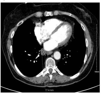
Further investigation with chest computed tomography angiogram was performed, which revealed the presence of a stable pericardial cyst measuring 22 x 17 mm at the level of the left atrium. Additionally, a low attenuating lymph node adjacent to the left atrium and bibasilar atelectasis was observed. These findings strongly indicate the presence of a benign pericardial cyst. (Figure 2)

Figure 1: EKG without any abnormalities detected.

Figure 2: CT chest showing pericardial cyst measuring 22 x 17 mm.
Discussion
Clinical Manifestation
Although the pericardial cyst is considered rare, it is the third most common type of mediastinal mass, comprising 33% [2]. Parmar et al. reported that pericardial cysts account for 17% of all mediastinum cysts, with a predominant location on the right side of the chest, where 75% appear at the right cardiophrenic triangle, while 22% of the cysts are located on the left side, and the remaining percentage in other mediastinum portions [3]. Some data show the average diameter of pericardial cysts is approximately 5.4 cm. However, the size of these cysts could be up to 13 x 8 x 5 cm, which is described in Manouchehr's case presentation [4]. Patients with these cysts are usually asymptomatic, 25% manifest symptoms of chest pain, dyspnea, consistent cough, retrosternal pressure, dysrhythmias, palpitations, or recurrent respiratory infections [8, 9]. Although most of the cysts are considered benign, they can cause several severe complications that might be life-threatening depending on their size and location. Ruptured cysts can cause cardiac tamponade, hemorrhage, or sudden cardiac death [5]. They can compress adjacent structures causing right ventricular obstruction flow, superior vena cava, or bronchial obstruction.
Diagnostic Methods
Since most pericardial cysts are asymptomatic, this condition is typically identified incidentally through diagnostic imaging. Chest X-ray, CT scan, MRI, and TEE are available to establish the diagnosis [3]. While pericardial cysts are often detected by chance by chest X-ray, CT scan is considered the most reliable option due to its ability to provide detailed information about the cysts [3]. The appearance of a thin-walled, well-defined mass without any septations or solid components on CT scan is characteristic of pericardial cysts. In some cases, elevated protein within the cysts can result in inconsistent density on CT scans making it difficult to differentiate from hematoma or neoplasm. The application of a diffusion-weighted technique using MRI to calculate apparent diffusion coefficients has the potential to address these challenges [10]. Despite the ability to provide excellent tissue structure and high accuracy, limitations in terms of cost and time pose obstacles to using MRI to routinely diagnose this condition [3]. Although commonly used as the initial approach in symptomatic patients, TEE has limited sensitivity for detecting pericardial cysts due to its restricted field of view and narrow window. According to Alkharabsheh’s study, TEE’s sensitivity in detecting pericardial cysts is 38% [11]. The accuracy of echocardiography may also be affected by patient factors such as obesity, obstructive pulmonary diseases, or previous surgery. Therefore, the American Society of Echocardiography Clinical recommends obtaining CT scans or MRI on suspected pericardial cysts image on chest X-ray or echocardiogram [12]. In cases where cysts are located at unusual sites, a combination of imaging modalities may be necessary for an accurate diagnosis.
Available treatments and Follow-ups
When selecting treatments for cysts, it is essential to adopt a systematic approach. Several treatment methods are available, such as conservation and follow-up, percutaneous aspiration, ethanol sclerosis, or cyst removal [3]. According to many researchers, the choice of treatment depends on the presence of symptoms, the size and location of the mass, and the patient's quality of life. Conservative management and regular follow-up are usually recommended in patients with small cysts and asymptomatic. Among the methods of follow-up, echocardiography is preferred because of its low risk of radiation compared to CTA. Based on the findings of Alkharabsheh's study, among 29 asymptomatic patients under conservative management with 23 months of periodic imaging follow-up, 48% of patients showed no change in cyst size, 33% experienced a significant decrease in diameter, and 17% exhibited a significant increase in maximum diameter [11]. However, periodic follow-ups may lead to patient anxiety and impose a burden in terms of treatment time and cost. Therefore, the European Society of Cardiology recommends using percutaneous aspiration and ethanol sclerosis as the first line of management for symptomatic and complex cysts, although the recurrence rate may be up to 30% [6]. Cyst removal could be done through an assisted video thoracotomy or surgery. This method is considered a second line of management of large cysts, symptomatic cases, or a prevention method when a patient has a significant risk of developing life-threatening conditions such as cardiac tamponade, sudden cardiac death, or airway obstruction [3]. However, surgical removal of cysts can pose complications related to anesthesia, cardiac dysrhythmias, such as supraventricular arrhythmia, or substantial bleeding resulting from erosion of the right ventricular wall [14, 15].
Case-Specific Features
The patient presented in this case report was incidentally found to have a pericardial cyst when she had the chest CTA done. Later on, she developed chest discomfort in the anterior chest without radiation, and without being related to exertion or precipitating factors. This pain might come from the pericardial cyst, however, since this patient has risk factors for cardiovascular disease such as hyperlipidemia, age, and remote smoking, a cardiac investigation for coronary artery disease is necessary. The result of the combination of EKG, echocardiography, and nuclear stress test showed no evidence of cardiac ischemia. The specificity and sensitivity of the nuclear stress test are 82% and 76%, respectively, and the likelihood of myocardial infarction in the next 2 years in a negative stress test is less than 1% [7]. Hence, the patient’s chest discomfort is likely not from coronary artery disease.
During follow-up visits, the patient had been experiencing anxiety and consistent concerns about the cyst, which may happen when choosing conservative management and regular follow-up, as previously discussed. In such situations, treatment options such as aspiration and ethanol sclerosis may be considered to alleviate patient stress. However, in our case, we chose to discuss and explain the likelihood of dangerous events or malignancy associated with the cyst and reassured the patient that the cyst carries minimal harmful risk. As a result, the patient comprehended the explanation and experienced significant relief. Therefore, in conservative management, it is crucial to provide reassurance and answer questions thoroughly to alleviate patient stress.
Conclusion
This case emphasizes the significance of considering pericardial cysts as a possible etiology for chest pain in patients with relevant symptoms. Diagnostic imaging modalities, including chest CT scan and TTE, play a pivotal role in establishing an accurate diagnosis. Surgical excision is a recommended treatment approach for large symptomatic pericardial cysts, whereas conservative management with periodic imaging surveillance may be appropriate for small symptomatic cysts. Long-term follow-up is crucial to monitor patient recovery, assess for potential recurrence, and detect any complications that may arise
References
- Qamar Y, Gulzar M, Qamar A, Sabry H, Minhas T. An Incidental Finding of a Large Pericardial Cyst. Cureus. 2022.
- Khayata M, Alkharabsheh S, Shah NP, Klein AL. Pericardial Cysts: a Contemporary Comprehensive Review. Current Cardiology Reports. 2019; 21(7): 64
- Kar SK, Ganguly T. Current concepts of diagnosis and management of pericardial cysts. Indian Heart Journal. 2017; 69(3): 364-70.
- Hekmat M, Ghaderi H, Tatari H, Arjmand Shabestari A, Mirjafari SA. Giant Pericardial Cyst: A Case Report and Review of Literature. Iranian Journal of Radiology. 2016; 13(1).
- Shiraishi I, Yamagishi M, Ayumi Kawakita, Yamamoto Y, Kenji Hamaoka. Acute Cardiac Tamponade Caused by Massive Hemorrhage from Pericardial Cyst. Circulation. 2000; 101(19).
- Maisch B, Seferović PM, Ristić AD, Erbel R, Rienmüller R. Guidelines on the Diagnosis and Management of Pericardial Diseases Executive SummaryThe Task Force on the Diagnosis and Management of Pericardial Diseases of the European Society of Cardiology. European Heart Journal. 2004; 25(7): 587-610.
- Matta M, Harb SC, Cremer P, Hachamovitch R, Ayoub C. Stress testing and noninvasive coronary imaging: What’s the best test for my patient? Cleveland Clinic Journal of Medicine. 2021; 88(9): 502-15.
- Chihara R, Force SD. Laparoscopic Approach for Treatment of Symptomatic Intra-Pericardial Cyst. The Annals of Thoracic Surgery. 2017.
- Najib MQ, Chaliki HP, Raizada A, Ganji JL, Panse PM, Click RL. Symptomatic pericardial cyst: a case series. European Journal of Echocardiography: The Journal of the Working Group on Echocardiography of the European Society of Cardiology. 2011; 12(11): E43.
- Raja A, Walker JR, Sud M, Du J, Zeglinski MR, Czarnecki A, et al. Diagnosis of pericardial cysts using diffusion weighted magnetic resonance imaging: A case series. Journal of Medical Case Reports. 2011; 5(1).
- Alkharabsheh S, Gentry III JL, Khayata M, Gupta N, Schoenhagen P, Flamm S, et al. Clinical Features, Natural History, and Management of Pericardial Cysts. The American Journal of Cardiology. 2019; 123(1): 159-63.
- Klein AL, Abbara S, Agler DA, Appleton CP, Asher CR, Hoit B, et al. American Society of Echocardiography Clinical Recommendations for Multimodality Cardiovascular Imaging of Patients with Pericardial Disease: Endorsed by the Society for Cardiovascular Magnetic Resonance and Society of Cardiovascular Computed Tomography. Journal of the American Society of Echocardiography. 2013; 26(9): 965-1012.e15.
- Meredith A, Zazai IK, Kyriakopoulos C. Pericardial Cyst. PubMed. Treasure Island (FL): StatPearls Publishing; 2022.
- Generali T, Garatti A, Piervincenzo Gagliardotto, Alessandro Frigiola. Right mesothelial pericardial cyst determining intractable atrial arrhythmias. Interactive Cardiovascular and Thoracic Surgery. 2011; 12(5): 837-9.
- Kaul P, Kalyana Javangula, S. Farook. Massive benign pericardial cyst presenting with simultaneous superior vena cava and middle lobe syndromes. Journal of Cardiothoracic Surgery. 2008; 3(1).

