Clinical Image - Volume 3 - Issue 6
The impact of trauma on the nevus: What dermoscopy reveals
Ghita Sqalli Houssini1*; Meryem Soughi1; Zakia Douhi1; Sara Elloudi1; Hannane Baybay1; Laila Tahiri Elousrouti2; Fatima Zahra Mernissi1
1Department of Dermatology, University Hospital Hassan II, Fez, Morocco.
2Anatomical pathology laboratory, University Hospital Hassan II, Fez, Morocco.
Received Date : Nov 01, 2023
Accepted Date : Nov 20, 2023
Published Date: Nov 27, 2023
Copyright:© Sqalli Houssini Ghita 2023
*Corresponding Author : Sqalli Houssini Ghita, Department of Dermatology, University Hospital Hassan II, Fes, Morocco.
Email: gsqallihoussini@gmail.com
DOI: Doi.org/10.55920/2771-019X/1587
Clinical Image
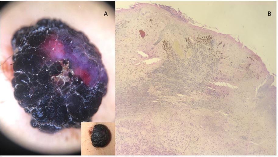
A 37-year-old female, devoid of prior pathologic history, exhibiting a brown tumor on her back for many years. Over the past year, the tumor grew larger, turning dark, becoming painful, and prone to bleeding. She reported occasional traumas during bathing and dressing. Clinical examination unveiled a 3cm tumor with a rough surface and a sessile base, encircled by peripheral bleeding. Upon dermoscopic examination, circular movements on the lesion revealed the tumor’s mobility, known as the “dimpling” sign. Furthermore, we observed the emergence of black dots and a central black, pavement-like area with reddish-purple hemorrhagic suffusion.Considering these findings, we initially contemplated traumatized dermal naevus, traumatized seborrheic keratosis, or solitary angiokeratoma. Histology depicted a largely ulcerated surface epidermis with numerous hair follicles. The dermis exhibited a benign tumor proliferation in nests of rounded naevus cells, accompanied by congestive and inflammatory changes, resulting in a diagnosis of traumatized dermal naevus Trauma affecting naevi can evoke patient anxiety due to alterations in their appearance. Conversely, architectural changes can complicate dermoscopic interpretation[1].Specific histological characteristics commonly found in traumatized nevi can make histological interpretation challenging, potentially leading to an overdiagnosis of melanoma, particularly when essential clinical information such as a history of trauma is unavailable [1].This emphasizes the need for a clinical, dermoscopic, and histological correlation. Our case demonstrates that dermoscopic signs, such as the pavement-like appearance, emergence of hairs, and hemorrhagic suffusion, align perfectly with histological findings. The “dimpling” sign observed through dermoscopy already points to the naevus nature of the tumor.
Conflicts of interest: The authors do not declare any conflict of interest.
Consent: The examination of this patient was conducted according to the Declaration of Helsinki principles.

Figure 1: (A) Dermoscopic appearance of a dark back tumor: emerging hairs, blackish pavement-like pattern, reddish-purple hemorrhagic suffusion and (B) Histological image shows a skin surface lined by a widely ulcerated epidermis with the presence of numerous hair shafts. The dermis shows a benign tumor proliferation arranged in nests and sheaths composed of nevus cells.
References
- Selan, Maruša, Daja Šekoranja, Mateja Starbek Zorko. A Traumat ized Melanocytic Nevus with Atypical Clinical and Dermoscopic Features: A Case Report and Review of the Literature. Acta Dermatovenerologica Alpina Pannonica et Adriatica. 2021; 30: 49-51.

