Clinical Image - Volume 3 - Issue 6
Multiple myeloma and exophthalmos
Soukaina El Amrani*; Kessi Eric Michel Charlemagne Junior; Amal Lahfidi; A.El Khamlichi; Firdaous Touarsa; Najwa El Kettani; Meriem Fikri; Mohamed Jiddane
Neuroradiology Department, Specialty Hospital of Rabat, Mohammed V University, Rabat, Morocco.
Received Date : Nov 04, 2023
Accepted Date : Nov 21, 2023
Published Date: Nov 28, 2023
Copyright:© El Amrani Soukaina 2023
*Corresponding Author : El Amrani Soukaina, Neuroradiology Department, Specialty Hospital of Rabat, Mohammed V University, Rabat, Morocco.
Email: Drsoukainael@gmail.com
DOI: Doi.org/10.55920/2771-019X/1588
Keywords: Multiple myeloma, orbital involvement, Exophthalmos.
Clinical Image
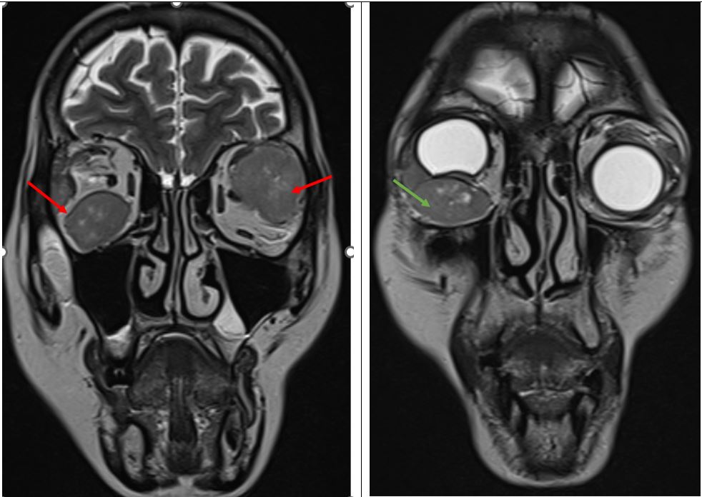
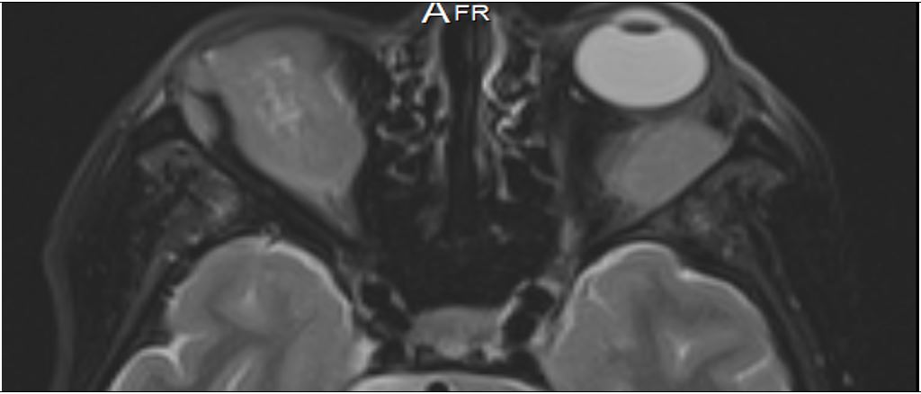
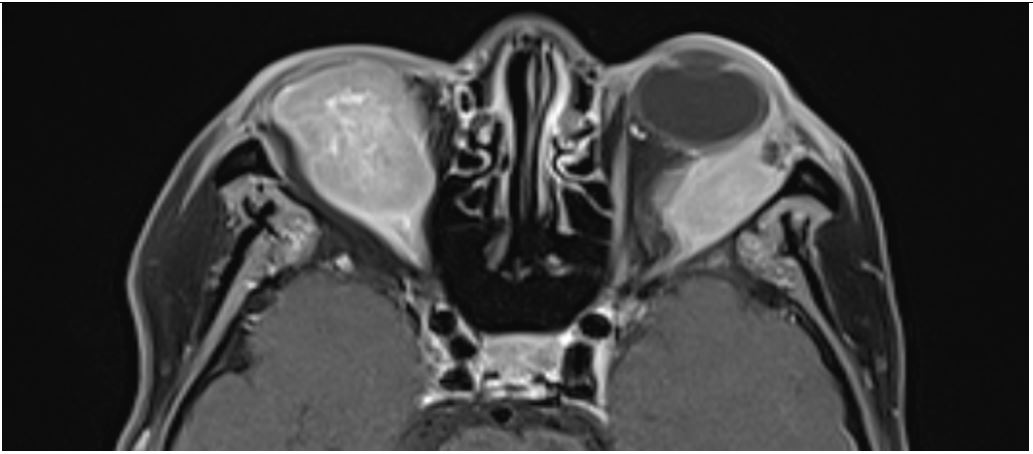
Extramedullary myeloma is a type of MM defined by the presence of extraskeletal (soft tissue or visceral) clonal plasma cells infiltrates. Orbital involvement is very rare and has been reported to involve conjunctiva, cornea, retina, orbital bones, and rectus muscles [1]. Orbital involvement presents as a solitary soft-tissue tumor originating from bone deposits and may be associated with no bone destruction. Orbital involvement is more frequently extraconal (90%) and located in the superior-temporal quadrant (75%); the involvement of recti muscles is very rare [2]. On CT, orbital myeloma is slightly dense than muscle, and contrast enhancement is slight and homogenous sometimes with dark zones representing spontaneous intralesional hemorrhage. On MRI, the lesion is isointense on T1 and has a moderate signal on T2. There is a significant contrast enhancement with central inhomogeneity; there is no diffusion restriction. Orbital MM is a very unusual cause of an orbital mass and can raise difficulties in differential diagnosis between lymphoma, metastases ,rhambdomyosracoma. Treatment of the orbital plasmacytoma is not well defined, includes local excision as a salvage surgery, exenteration or radiotherapy and additional chemotherapy. Its appearance is associated with an advanced stage of multiple myeloma and a poor prognosis. We report the case of a 60-year-old woman with known mult iple myeloma presented with bilateral painful proptosis, right conjunctival chemosis and a right palpable mass. On examinat ion, it was found that she has bilateral exophthalmos more marked on the left side and decreasing mobility in abduction. A magnetic resonance imaging showed multiples large extraconal masses each involving the lateral recti muscles especially on the left (Fig.1), scalloping on the eyeballs and causing exophthalmos grade 4 (Fig.2), heterogeneously enhancing on contrast images (Fig.3). There was no bony orbital lysis. Shortly thereafter, the patient expired.
Declaration of interests: The authors declare that they have

Figure 1: Coronal T1 MRI showing isointense bilateral masses involving of both lateral rectus muscles(red arrow) and right inferior rectus muscle causing indentation of globe and proptosis (green arrow).

Figure 2: Axial T1 Weighted images showing extraconal masses causing exophtalmos grade 4.

Figure 3: Axial T1 Weighted fat-saturated T1-weighted imaging with injection: There is a heterogeneous enhancement of the intral orbital masses.
no known competing financial interests or personal relationships that could have appeared to influence the work reported in this paper.
Patient consent: Written informed consent for publication was obtained from patient.
References
- Thoumazet F, Donnio A, Ayeboua L, Brebion A, Diedhou A, Merle H. Orbital and muscle involvement in multiple myeloma. Can J Ophthalmol. 2006; 41(6): 733-6.
- Saffra N, Gorgani F, Panasci D, Kirsch D. Diplopia and proptosis due to isolated lateral rectus plasmacytoma in a patient with multiple myeloma. BMJ Case Rep. 2019; 12(7): e229178.

