ISSN Number
ISSN 2771-019X-

-

Impact Factor
1.2*
ISSN Number
ISSN 2771-019X
Impact Factor
1.2*1National Capital Consortium, Department of Internal Medicine, Walter Reed National Military Medical Center, Bethesda, MD 20889, USA.
2Department of Gastroenterology, Walter Reed National Military Medical Center, Bethesda, MD 20889, USA.
National Capital Consortium, Department of Internal Medicine, Walter Reed National Military Medical Center, Bethesda, MD 20889, USA.
Email: madisonperegoy1@gmail.com
Received : Oct 05, 2024,
Accepted : Nov 08, 2024
Published : Nov 15, 2024,
Archived : www.jclinmedcasereports.com
Copy right Statement: Content published in the journal follows Creative Commons Attribution License (http://creativecommons.org/licenses/by/4.0). © Peregoy M (2024).
Journal: Open Journal of Clinical and Medical Case Reports is an international, open access, peer reviewed Journal mainly focused exclusively on the medical and clinical case reports.
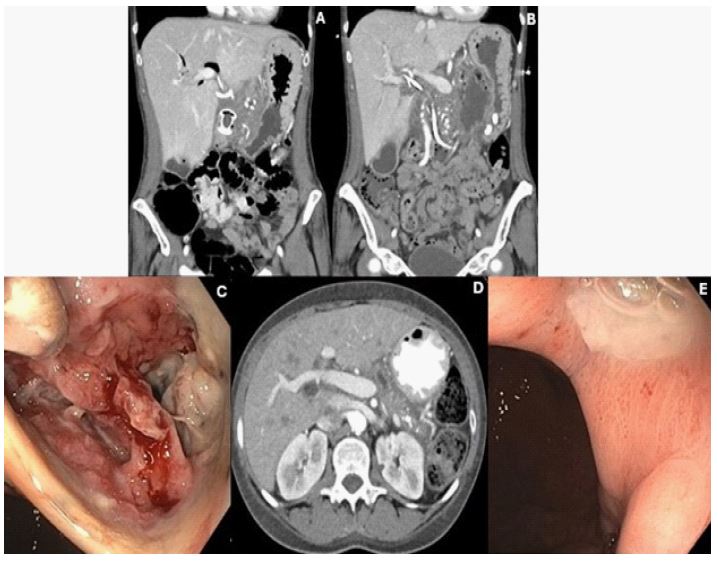
A 55-year-old female with a history of chronic alcohol-related pancreatitis presented with abdominal pain and anorexia. Cross-sectional imaging revealed a homogenous large fluid collection located in the pancreatic head consistent with pseudocyst, which was followed with repeat imaging studies. She subsequently underwent uncomplicated Endoscopic Ultrasound (EUS) guided cystoduodenostomy with placement of a lumen opposing metal stent along with common bile duct stent placement for biliary obstruction (Figure 1A). One month later she represented with recurrent abdominal pain, anorexia, fever, and leukocytosis. Repeat imaging studies revealed a new complex peripancreatic fluid and gas collection concerning an infected acute pancreatic fluid collection (Figure 1B). She was scheduled for EUS- guided drainage the following day and interestingly improved symptomatically overnight just prior to the procedure. Upper endoscopy the following day revealed a 4 cm defect in the posterior gastric wall filled with healthy-appearing granulation tissue and necrotic debris, indicative of spontaneous decompression of the infected pancreatic fluid collection (Figure 1C). CT imaging revealed resolution of this fluid collection following this spontaneous decompression (Figure 1D). Acid suppression therapy was discontinued and interval endoscopy after 3 weeks showed resolution of the defect (Figure 1E).
Financial or competing interests disclosure: There are no actual or potential conflicts to disclose.
