ISSN Number
ISSN 2771-019X-

-

Impact Factor
1.2*
ISSN Number
ISSN 2771-019X
Impact Factor
1.2*Department of Breast Surgery, West China Hospital, Sichuan University, Chengdu, Sichuan Province, China.
Department of Breast Surgery, West China Hospital, Sichuan University, Chengdu, Sichuan Province, China.
Email: 29247901@qq.com
Received : Nov 06, 2024,
Accepted : Dec 05, 2024
Published : Dec 12, 2024,
Archived : www.jclinmedcasereports.com
Copy right Statement: Content published in the journal follows Creative Commons Attribution License (http://creativecommons.org/licenses/by/4.0). © Chen J (2024).
Journal: The Journal of Clinical and Medical Images, Case Reports (JCMICR) is a fantastic resource for keeping up with the latest clinical advancements and for publishing case reports and clinical images related to a variety of medical illnesses.
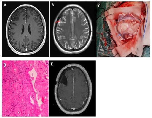
A 32-year-old woman with hypertension was referred to neurosurgery department with 3-day history of drooping of left side of mouth. She has been taking telmisartan for hypertension for five years. Magnetic resonance imaging (MRI) of the brain showed neither infarction nor hematoma, but a ill-defined lesion without enhancement, highly suspected to be a low-grade glioma, was found in the right frontal lobe on T1-weighted images (Figure 1A, white arrow) and T2-weighted images (Figure 1B, red arrow).
Although the intracranial lesion’s imaging characteristics were very similar to that of a low-grade glioma preoperatively, we were also not 100% sure about that the lesion was a low-grade glioma. Just in case that there was another exception, and the patient just only presented with drooping of left side of mouth ,the symptom of Bell’s palsy, she had no any other symptoms related to increased intracranial pressure, such as headache, nausea, vomiting, as well as epilepsy. So we recommended follow-up visit first, because even it was a low-grade glioma, it progressed very slowly, we can wait and see, but the patient and her family asked for surgery after discussing with each other.
Then the tumor-like lesion was removed subsequently, and it was found to be milk-white, diffuse and hard during the operation (Figure 1C, blue arrow). The pathological result (Figure 1D, Hematoxcylin eosin staining) revealed interstitial fibrous hyperplasia, hyaline degeneration and thickening vessel wall in the lesion tissue. Based on the pathological findings, combining with the patient’s 5-year history of hypertension, the pathologists concluded that the lesion was caused by the chronic hypertension. And there was no molecular detection supporting for the diagnosis of tumor. At 6 months of follow-up, a MRI scan showed no recurrence in the resection cavity (Figure 1E).