ISSN Number
ISSN 2771-019X-

-

Impact Factor
1.2*
ISSN Number
ISSN 2771-019X
Impact Factor
1.2*1Khwarizmi Institute of science and technology, Qeshm Island, Hormozgan, Iran.
2Congress 60 Non-governmental organization, Iran.
Khwarizmi Institute of science and technology, Qeshm Island, Hormozgan, Iran.
Email: arvinland@yahoo.com
Received : Nov 23, 2024,
Accepted : Dec 19, 2024
Published : Dec 26, 2024,
Archived : www.jclinmedcasereports.com
Breast cancer is the most prevalent cancer in women and breast cancer metastasis is one of the main mortal causes of women worldwide. Opium has been used as a pain killer for cancer patients. The present short report is the early results of the study aimed to evaluate the gene expression profiling of the rat models of breast cancer under a new taper-up-off treatment method with opium tincture.
The present study included Wistar rat models of breast cancer distributed to four groups of taper-up-off treatment with different dosages of opium tincture, a group of breast cancer models with no treatment, a sham group, and a non-cancer normal control. Mammary tissues were collected, and after the RNA extraction, whole-genome expression profiling was performed, and results were confirmed by Real-time PCR. Pathway analysis was conducted on expression results.
Gene expression profiling showed hundreds of genes that were involved in tumorigenesis, immune system, cell proliferation, and most significantly metastasis pathways (including OPG/RANK/RANKL, Transforming growth factor-β, Wnt, and JAK-STAT signaling pathways) were up-regulated in breast cancer models and down-regulated after treatment with opium tincture, suggesting potential metastasis inhibition effect of taper up-off treatment method of opium in breast cancer cells.
Early findings of the present study determined several pathway alterations, especially a potential metastasis inhibition effect of opium consumption. Results may shed light on possible opportunities for the development of new treatments for breast cancer metastasis.
Keywords: Breast cancer; Opium; Taper up-off; Microarray; Metastasis.
Copy right Statement: Content published in the journal follows Creative Commons Attribution License (http://creativecommons.org/licenses/by/4.0). © Haghighatfard A (2024).
Journal: The Journal of Clinical and Medical Images, Case Reports (JCMICR) is a fantastic resource for keeping up with the latest clinical advancements and for publishing case reports and clinical images related to a variety of medical illnesses.
Breast cancer is cancer that develops from breast tissue, especially in cells from the lining of milk ducts and the lobules and its symptoms include lumps in the breast, alterations in the shapes of breasts, dimpling of the skin, fluid coming from the nipple and red or scaly patch of skin and in some cases bone pain, swollen lymph nodes and shortness of breath [1,2].
Obesity, lack of physical exercise, alcohol consumption, hormone replacement therapy especially during menopause, and a family history of breast cancer are the main risk factors for developing breast cancer [3,4]. Genetic mutations including mutations in the BRCA gene are associated with inherited breast cancer [5].
Epidemiological studies show that the prevalence of breast cancer has dramatically increased globally during the last frothy years and statistics estimate that breast cancer is the cause of about 43000 deaths in the United States each year [6].
Opium is derived from the opium poppy (Papaver somniferum L.) and is one of the earliest plants recorded for medicinal use and is a dried latex, obtained from the seed capsules. Opium has been used as the main source of Morphine based pain killers [7]. The opium poppy is well known as a double-edged sword in the history of medicine which on one hand works as a source for pharmaceutical products of alkaloid-based pain relievers, addiction treatment [8], and even potential cancer treatment medicine [9], and in the other hand driven the most prevalent epidemic addiction of human history [10].
Opium effects are not limited to the opioid receptors and Central Nervous System (CNS) and it has been reported that the immune system, kidney functions, and respiratory and cardiovascular systems could be affected by opium consumption [11,12].
There is a great lack of knowledge about the opium effects on the gene expression and epigenetic patterns of mammalian genes. While for decades several opioid-based agents have had a major role in the pain reduction of several patients including cancer patients, the role of opiate alkaloids on epigenetics, biological processes, and metabolism of cancer cells, is not clarified. Limited studies about the potential tumor-promoting, proliferation, and migration effects of opiates are not conclusive, and both growth-promoting and anti-tumor effects have been reported in these studies [13]. Developing new molecular genetic techniques such as microarray and RNA sequencing helped us to shed light on the complexity of the role of opium consumption in cancer treatment processes aimed to draw a holistic picture showing the whole effects of opium on the gene expression pattern of cancer cells as well as all whole body of the patient.
In the present study effect of opium tincture taper up-off treatment in a laboratory-designed method on the expression profile of rat models of breast cancer was evaluated. We aimed to understand how the oral consumption of opium in a bell-shaped dose curve could alter several gene expression patterns that were previously affected by breast cancer.
Animal modeling of breast cancer
Rodents, especially mice and rats, are the most popular animals for breast cancer research. In the present study, we obobtained female Wistar albino rats at the age of 35-40 days old, from the animal laboratory of Pasture Institute of Iran, Tehran, Iran. All rats were placed in an air-conditioned room and inhabited in polycarbonate cages lined with wood chip bedding, under a 12 hr. light/dark cycle and room temperature of about 25°C throughout the experiment in the animal room. Due to the situation adaptation, all rats were maintained on a standard pellet diet and tap water for two weeks before starting the experiment. All experiments were carried out by the ARRIVE guidelines (Animal Research: Reporting of in Vivo Experiments) checklist and the ethical standards approved by the local ethical committee. A single oral dose of Dimethylbenz (a) anthracene (DMBA) solution dissolved in sesame oil (100 mg/kg) was used to induce mammary gland tumors in female rats aged 45-50 days. The dose of DMBA was selected according to the previous studies [14]. The rats were randomly divided into seven groups and each group included twelve female rats (aged 45-50 days and weighing 200-250 grams). During the animal modeling, rats of the control group were given a daily dose of distilled water orally (5 mL/kg b.wt) for 22 weeks, with a single dose of sesame oil (5 mL/kg b.wt) after one week. Breast cancer rat models were given a daily oral dose of distilled water for 22 weeks, with a single oral dose of DMBA dissolved in olive oil (100 mg/kg b.wt) after one week to induce breast cancer. The list of groups and descriptions of each group are presented in Table 1.
Taper up-off treatment with opium tincture
Force-feeding of rats by opium tincture had been performed based on a novel laboratory-designed method of taper-up-off treatment called the Dezhakam-Step-Time (DST) method. The opium tincture dosage starts from the lowest dose, and the dose increases at a 20% rate each day, (or 0.8 coefficient) for each oral feeding (two times per day), until 18 oral feeding (9 days). Then the dosage of feeding is reduced by the same 20% rate (or 0.8 coefficient) for the next 18 oral feeding (9 days). The rodent models received the same dosage in the first time and last time of oral feeding. Four sets of dosages were used in four protocols called “DST1”, “DST2”, “DST3”, and “DST4” each protocol used in one experiment group (Table 1). In the “DST1” protocol, the opium tincture dosage started at 2.35 mg/kg force-feeding and increased with 0.8 coefficient until 65.58 mg/kg in the 18th oral feeding time. “DST2”, “DST3”, and “DST4” started at 3.52 mg/kg, 4.7 mg/kg, and 5.87 mg/kg respectively.
Scarification and tissue preparation
After the treatments, all the rats on the same day were sacrificed at two similar standard animal laboratories using the diethyl ether and a cervical dislocation to collect mammary tissues. All euthanasia processes were monitored by the researchers until procedures were accomplished due to confirmation that all procedures were performed based on the ARRIVE55 Guidelines checklist and standard protocols [15,16]. Extracted mammary glands were dehydrated and immediately washed with ice-cold normal saline frizzed in liquid nitrogen at -80°C for further processes.
RNA extraction
Tissue samples were put in the process of RNA extraction. After the unfreezing of the tissue samples, RNA was extracted from mammary glands according to standard protocols using by RNA Purification kit (Gene JET™ RNA Purification Kit#K0732, Thermo Scientific - Fermentas, Latvia). Due to the removal of contamination of genomic DNA out of extracted RNA, the DNase Treatment & Removal Reagents (DNase I, RNase-free (#EN0521) Thermo scientific - Fermentas, Latvia), were used according to the manufacturer’s protocol. To calculate extracted RNAs quantity the absorbance of samples in 230, 260, and 280 nanometer UV waves was examined using Nanodrop-1000 equipment for UV-spectroscopy. Also for the evaluation of extracted RNA quality, gel electrophoresis with one percent agarose gel was performed for all samples. Due to the minimum requirements of quality and integrity of extracted RNAs for microarray analysis, RNA integrity values (RIN value) were evaluated for all samples by Agilent 2100 Bioanalyzer (Agilent Technologies).
| No. | Group name | Description of group |
|---|---|---|
| 1 | A | Rat modelof breast cancerwith no treatment |
| 2 | B | Rat modelof breast cancerwith DST1 treatment |
| 3 | C | Rat modelof breast cancerwith DST2 treatment |
| 4 | D | Rat modelof breast cancerwith DST3 treatment |
| 5 | E | Rat modelof breast cancerwith DST4 treatment |
| 6 | F | Rat modelof breast cancertreated with sham |
| 7 | G | Control groupincluded normal rats with no cure |
DST1: Dezhakam-step-time treatment method with 2.35 mg/kg force-feeding of opium tincture as first dosage, DST2: Dezhakam-step-time treatment method with 3.52 mg/kg force-feeding of opium tincture as first dosage, DST3: Dezhakam-step-time treatment method with 4.7 mg/kg force-feeding of opium tincture as first dosage, DST4: Dezhakam-step-time treatment method with 5.87 mg/kg force-feeding of opium tincture as first dosage.
| No. | Groups compared withG group | Total DEGs | Up-regulated genes | Down-regulated genes |
|---|---|---|---|---|
| 1 | A | 2606 | 1560 | 1046 |
| 2 | B | 1849 | 1109 | 740 |
| 3 | C | 1820 | 1066 | 754 |
| 4 | D | 1219 | 644 | 575 |
| 5 | E | 1035 | 517 | 518 |
| 6 | F | 2733 | 1679 | 1054 |
(A) rat model of breast cancer with no treatment, (B) rat model of breast cancer with DST1 treatment, (C) rat model of breast cancer with DST2 treatment, (D) rat model of breast cancer with DST3 treatment, (E) rat model of breast cancer with DST4 treatment, (F) rat model of breast cancer with sham treatment, (G) The Control group included normal rats with no cure.
| Number | Group comparisons | Number of significantlyaltered Pathways |
|---|---|---|
| 1 | A vs. G | 458 |
| 2 | B vs. G | 394 |
| 3 | C vs. G | 352 |
| 4 | D vs. G | 236 |
| 5 | E vs. G | 204 |
| 6 | A vs. F | 11 |
| 7 | F vs. G | 466 |
(A) rat model of breast cancer with no treatment, (B) rat model of breast cancer with DST1 treatment, (C) rat model of breast cancer with DST2 treatment, (D) rat model of breast cancer with DST3 treatment, (E) rat model of breast cancer with DST4 treatment, (F) rat model of breast cancer with sham treatment, (G) The Control group included normal rats with no cure.
CDNA synthesis
Copied DNA synthesis was performed using Transcription First Strand cDNA Synthesis Kit (Revert Aid Premium First Strand cDNA Synthesis Kit #K1652, Thermo scientific -Fermentas, Latvia) protocol and Applied Biosystems 9800 FAST Thermal Cycler instrument.
DNA microarray testing’s
Gene expression profiling was conducted using the Affymetrix GeneChip™ Rat Genome 230 2.0 Array a whole-genome analysis tool aimed at expression study of oncogenes, toxicology, neurobiology, and other rat models. Labeling and fragmentation of aRNA targets, hybridization, and scanning were performed based on standard protocol (Affymetrix Santa Clara, CA). First, the Quality Control (QC) was evaluated by Agilent Bioanalyzer and then hundred nanograms of extracted RNAs of each sample were processed by the GeneChip 3′ IVT Express Kit and were reverse transcribed and converted to double-stranded cDNA before biotin labeling during transcription. The fragmented areas of the sample were hybridized on Affymetrix GeneChip™ Rat Genome 230 2.0 Array for sixteen hours at 45° Centigrade temperature. In the next step, arrays were washed and stained using the GeneChip Hybridization, Wash, and Stain Kit on the GeneChip Fluidics Station 450. At the final step, chips were scanned by the Affymetrix GeneChip Scanner 3000, and all arrays passed the QC criteria examination.
The GeneChip analysis was performed with Microarray Analysis Suite (MAS) 5.0, Data Mining Tool 2.0, Genesis 2.0 (GeneLogic Inc., Gaithersburg, MD, USA) and Microarray Database software (available at http://www.affymetrix.com). Normalization and scaling of all represented genes in the GeneChip were conducted to a hundred signal intensity. False-positive results were filtered by MAS 5.0 and passed genes were analyzed by Genesis 2.0 (GeneLogic Inc., Gaithersburg, MD, USA). Genes with a ratio greater than two-fold and significant adjusted P values (after Bonferroni testing of multiple comparison correction) were selected and listed as the differentially expressed gene.
Real-time PCR examinations
All significantly differentially expressed genes in microarray results had been confirmed with Real-time PCR. First, the serial dilutions (1:4) of pooled cDNA were obtained from RNA samples of randomly chosen normal rats and used to create the standard curves. Primers and probes for differentially expressed genes were designed using “MEGA7” software and were checked on the BLAST-Primer section of the NCBI website. Quality checking criteria of qPCR assessments included an R2 value that should be more than 0.99 in the standard curve and no signal detection for no-template control samples. The PCR reaction efficacy was assessed by Lin-Reg PCR (Amsterdam, Netherlands) online free software. Real-time PCR was performed by TaqMan® PCR Starter Kit (Thermo scientific - Fermentas, Latvia) and CFX96 Touch Real-Time PCR Detection System (BIO-RAD, California, United States) and then the ratio calculations were performed using the Pfaffle formula.
Gene ontology analysis and pathways based on expression data
Pathway-based enriched functional analysis and gene ontology analysis were conducted on the final list of differentially expressed genes (DEGs) that showed in microarray study and confirmed in Real-time PCR. The integrated enrichment algoalgorithms of online Database for Annotation, Visualization, and Integrated Discovery (DAVID 6.8 version) and free online DAVID software (Strand Genomics, Redwood City, CA, USA) are used for functional annotation and gene ontology analysis. For pathways-based assessments and gene ontology mapping of DEGs, an online database called Kyoto Encyclopedia of Genes and Genomic (KEGG) pathway enrichment tool was used. Visualization of group comparisons in DEGs, Gene ontologies, and KEGG pathways as Venn diagrams drawn with online software of Van de Peer Lab Bioinformatics and Evolutionary Genomics.
Statistical analysis
Statistical examinations were conducted by the 25th version of Statistical Package for Social Science (SPSS) software. First of all, the Kolmogorov-Smirnov test is used for variables normal distribution analysis. Next, One-way ANOVA test was used for statistical differences in multiple group comparisons. Covariates were including of RNA integrity number, cDNA synthesis quality, plates/runs of qPCR, and primer efficiency. Due to the control of any potential confusion, the persistence of the significant difference between groups was calculated by the ANCOVA test. Bonferroni correction was used for multiple comparison corrections and the descriptive data are expressed as mean ± SD.
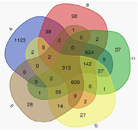
The mRNA level of all differentially expressed genes in microarray examinations was evaluated with Real-time PCR for confirmation. Findings determined that Real-time PCR results confirmed the results of microarray analysis in all comparisons between groups in both direction and level of expression alteration. Summary results of significantly differentiated genes revealed in the microarray tests (and confirmed in Real-time PCR) in each group had been presented in Table 2 and as Venn plots in Figure 1.
Enrichment pathway analysis showed several molecular pathways were altered in all breast cancer rat models with and without treatment. There were shared significantly altered DEGs in all groups under opium treatments. Pathways analysis showed opium treatment groups especially with “DST3” and “DST4” protocols had altered tumor suppressor genes and proto-oncogenes, transcription factors related to interleukin genes and metabolic pathways; immune system, natural killer cells regulators, and most significantly pathways related to metastasis inhibition.
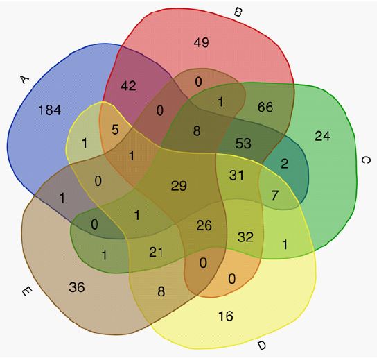
The most significantly altered pathways which their down regulations were shared between the four treatment groups were including OPG/RANK/RANKL signaling pathway (p=0.00005 in group B, p=0.00005 in group C, p=0.00004 in group D and p=0.00004 in group E), Transforming growth factor (TGF)-β signaling pathway (p=0.00003 in group B, p=0.00003 in group C, p=0.00003 in group D and p=0.00003 in group E), Wnt signaling pathway (p=0.00005 in group B, p=0.00005 in group C, p=0.00004 in group D and p=0.00004 in group E) and JAK-STAT signaling pathway (p=0.00005 in group B, p=0.00004 in group C, p=0.00003 in group D and p=0.00004 in group E). Statistical data of pathway analysis between treatment groups compared to controls are presented in Table 3 and as Venn plots in Figure 2. No significant DEGs were found between group A and group F (sham group).
Breast cancer is a lethal cancer in women worldwide which may lead to metastatic breast cancer and metastasize to lung, bone, liver, and even brain tissues [17,18]. Our gene expression findings showed that the new oral opium consumption could affect several molecular pathways associated with metastasis and potentially could reduce the rate of tumor progression and metastasis.
It has been determined that morphine and morphine-based medications could directly affect the central nervous system as a pain reliever, but their activities on tumor growth are still contradictory and evidence for both growth-promoting and growth-inhibiting effects has been reported [19]. It is important, that the ingredients of opium include about 50 alkaloids, which may affect molecular mechanisms and epigenetics patterns associated with the metastasis [20].
OPG/RANK/RANKL signaling pathway is highly expressed in breast cancer cells and leads to the directed migration of mammary epithelial cells [21] and in metastatic tumors, cells can directly secrete RANKL or induce the production of RANKL, which in turn causes a bone matrix degradation and dramatic increase of growth factors and cytokines, that leading tumor cells migration especially to bones [22].
Down-regulation of the OPG/RANK/RANKL signaling pathway in opium treatment groups may suggest a potential cell migration inhibition effect of opium tincture.
TGF-β signaling pathway plays an active role in the expression of cytokine as well as cellular functions, including cell cycle arrest, apoptosis, angiogenesis, and cell migration [23]. The upregulation of TGF-β signaling has been reported in breast cancer metastasis in bone tissue [24].
The Wnt signaling is one of the most conserved signaling pathways indicated as a key to controlling embryonic and organ development and its de-regulations could cause cancer progression and metastasis [25]. The Wnt signaling activities have a crucial role in breast cancer, such as immune microenvironment regulation, and therapeutic resistance [26]. The over-expression of nuclear β-Catenin as a pivotal component of the Wnt signaling pathway is important in tumorigenesis and metastasis, which is usually induced by aberrant Wnt activation [27]. Down expression of several genes in the Wnt signaling pathway including the β-Catenin gene (CTNNB1) detected in groups A, B, C, and D showed a potential effect of Wnt signaling inhibition of opium tincture.
The JAK/STAT signaling pathway has been involved in tumorigenesis, maintenance, and metastasis of breast cancer cells [28]. IL-6 mediated signaling pathway as a cytokine pathway, in turn, activates JAK/STAT signaling pathway. Interestingly active IL-6/JAK/STAT3 signaling would drive cancer cell proliferation, suppression of the anti-tumor immune response, and invasive metastasis [29]. Previous studies determined the potential immunosuppressive effects of opium on inflammatory mediators like C- Reactive Protein (CRP), interleukin-17, and interleukin-1 in the cardiovascular system [30]. Results of the present study showed dramatic downregulation of interleukin-6 (IL-6) in group C and group D that could lead to IL-6/JAK/STAT3 inhibition and metastasis inhibition as well.
Multiple mechanisms underlie breast cancer metastatic dissemination and it seems that the interpretation of opium’s effects on the metastasis signaling pathways is strongly dependent on the dosage and duration of consumption. While Opium has been prescribed as a painkiller for cancer patients in the metastasis phase, and it’s due to the analgesic, hypnotic, and antidiarrheal effects of opium, as a highly addictive substance [31] there is a huge obstacle to using opium as a potential treatment for cancer. It is highly essential to find a protocol that could prevent the dependency on opium and our newly presented taper-up-off protocol is aimed to achieve a potentially safe treatment protocol for opium consumption.
Limitations
We have reported the early results of gene expression profiling of breast cancer cells of rat models treated with a novel protocol of opium tincture consumption. To achieve certain conclusions, pathological analysis and longitudinal studies are needed. Additionally, there is a clinical difference between rodent breast cancer metastasis which usually occurs in the lung, with human breast cancer metastasize which usually happens in lymph nodes, liver, bone, and brain [32].
Our gene expression profiling findings may shed light on the potential effects of opium tincture treatment protocol to inhibit metastatic molecular activities in breast cancer cells. While opium is an addictive substance, it seems that if we could use oral opium tincture in a taper-up and taper-off protocol, there are some potential opportunities for advancing the therapy and the development of anti-metastasis treatments based on opium alkaloids’ effects.
Conflict of interest: There is no conflict of interest.
